







Technique is a tool. Anatomy is the map. Most injectors are taught the tool first and the map second, or not at all — they learn a set of injection points, they reproduce them competently, and they never develop a mental model of what is underneath those points. That works until it doesn't.
Facial vascular anatomy is the foundation of everything we do in aesthetics, and safety starts before the needle touches the face. Not when you aspirate. Not when you see blanching. Before. The moment you survey a face and decide where to place a product, you are either reasoning from a vascular map or you are not.
This page is that map for one vessel — the facial artery — and it is deliberately the first thing in this cluster because nearly every serious injectable complication of the lower two-thirds of the face traces back to it, its branches, or the vessels it talks to. Learn this one artery properly and the rest of facial vascular anatomy becomes a network you can extend, rather than a list you have to memorise.
Where the map begins: the carotid bifurcation
Start upstream, because the reason a complication in the lip can end in the eye only makes sense if you start upstream.
The common carotid artery bifurcates into the internal carotid and the external carotid. That single branch point is the most consequential fork in aesthetic medicine. The internal carotid supplies the brain and, by way of the ophthalmic artery, the eye. The external carotid supplies the face.
Two separate territories, on paper. In practice, they are joined across the midface and the glabella by a set of arterial connections that make the face one continuous, communicating vascular bed. That is the entire reason a filler embolus can leave the territory you injected and arrive somewhere you never touched.
The facial artery is a branch of the external carotid. It arises in the neck, runs deep and anteriorly, and then does something clinically important: it climbs over the inferior border of the mandible to enter the face.
Emergence at the antegonial notch
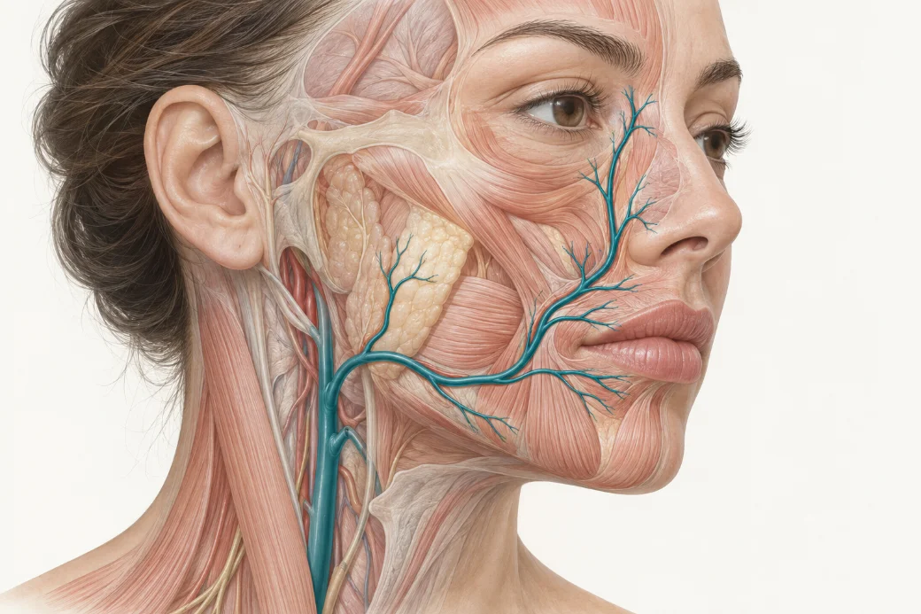
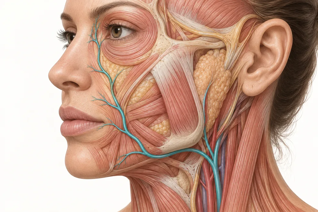
Lateral view. Find the masseter. Just anterior to the masseter's anterior border, along the inferior margin of the mandible, there is a small concavity in the bone — the antegonial notch. That is where the facial artery crosses from the neck into the face, and it is the one point on this vessel's entire course that you can reliably palpate. You can feel the pulse there on most patients.
Two things to hold onto about this landmark.
First, at this point the artery is deep. Pooled cadaveric and ultrasound data put the facial artery at roughly 6 mm from the skin surface at its mandibular origin — an average depth of 5.98 to 6.62 mm across a 2025 meta-analysis of twelve studies (Trzeciak et al., Aesthetic Plastic Surgery, 2025; doi:10.1007/s00266-025-04833-9). It is not a superficial vessel where it enters the face. It becomes one later.
Second, this is the practical starting point of your mental tracing. When I teach the facial artery, I don't start at the lips, because the lips are where the injecting happens and that biases the picture. I start at the notch, because from there the vessel has to travel to reach every structure you treat, and the route it takes is what puts it in your way.
The ascending course: notch to medial canthus
From the antegonial notch the facial artery runs superiorly and medially across the face, deep at first, becoming progressively more superficial as it ascends, and terminating at the medial canthus as the angular artery.
That is the sentence to memorise. Everything else on this page is detail hung on it.
The depth gradient matters as much as the route. The same meta-analysis reports mean depth rising from roughly 6 mm at the mandibular origin to 8.36–9.20 mm at the level of the oral commissure, and 9.52–10.51 mm at the level of the nasal ala (95% CI). But those means describe the main trunk in the deep plane — and by the time the vessel reaches the angular segment at the medial canthus, ultrasound work puts it at around 1.0 mm from the skin surface. An artery that started six millimetres down finishes a millimetre down. Depth is not a fixed property of a vessel. It is a property of a vessel at a location.
I want you visualising that ramp underneath the skin every time you survey a face.
The branches you meet along the way
Off that ascending trunk come the branches that supply almost everything an injector treats in the lower face.
Inferior labial artery
Arises from the facial artery near the oral commissure and runs medially to supply the lower lip. Present in the large majority of dissections, but not universal — a 102-hemiface cadaveric study found it in 78.43% of specimens (Nguyen et al., Archives of Craniofacial Surgery, 2024;25(2):77–84).
Superior labial artery
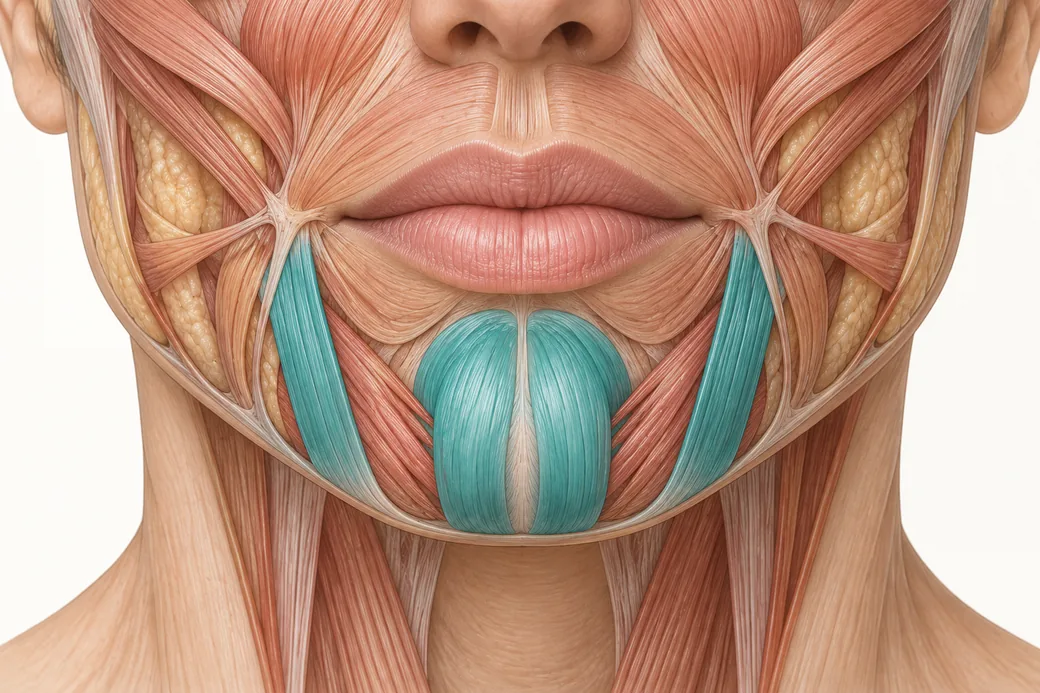
Arises above the commissure and runs medially to supply the upper lip. It is the more consistently present of the two labial arteries — 87.25% in the same series — and it is the vessel most directly implicated in lip complications. In vivo ultrasound of 41 volunteers put its mean depth in the upper lip at 5.6 ± 0.13 mm, with the artery sitting in the submucosal plane in 58.5% of measurements, intramuscular in 36.2%, and subcutaneous in only 5.3% (Cotofana et al., Aesthetic Surgery Journal, 2020;40(12):1327–1335). In 83% of upper lips it was found within the red lip rather than above the vermilion border.
The columellar branch
This is the one that surprises people. As the superior labial artery reaches the philtrum, it commonly gives off a columellar branch that ascends toward the nasal septum and base. Published series put the superior labial artery as the source of the columellar supply in roughly 72.5% of specimens. So there is a direct arterial road from the upper lip to the nose. Not a theoretical connection — a named branch.
This is why a patient treated in the lip and philtral column can call you about the nose.
Lateral nasal artery
Arises as the facial artery passes alongside the nasal ala and supplies the nasal sidewall, ala and dorsum. In the 2024 cadaveric series it was the single most common termination point of the facial artery — the vessel ended there in 32.35% of hemifaces.
Angular artery — the terminus
Where the facial artery continues past the ala, it is named the angular artery and it terminates in the medial canthus region. Combined across published series, termination as either the lateral nasal or the angular artery accounts for roughly 70% of facial arteries (Koziej et al., Clinical Anatomy, 2022).
The angular terminus is the single most important two centimetres of this map, and it gets its own section.
Why the medial canthus is the hinge point of the whole map
At the medial canthus, the angular artery arrives superficially — around 1 mm deep on ultrasound — and it arrives in a neighbourhood full of vessels that do not belong to the external carotid at all.
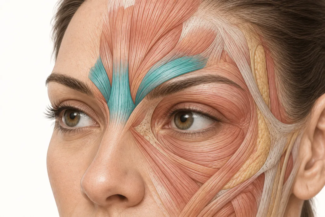
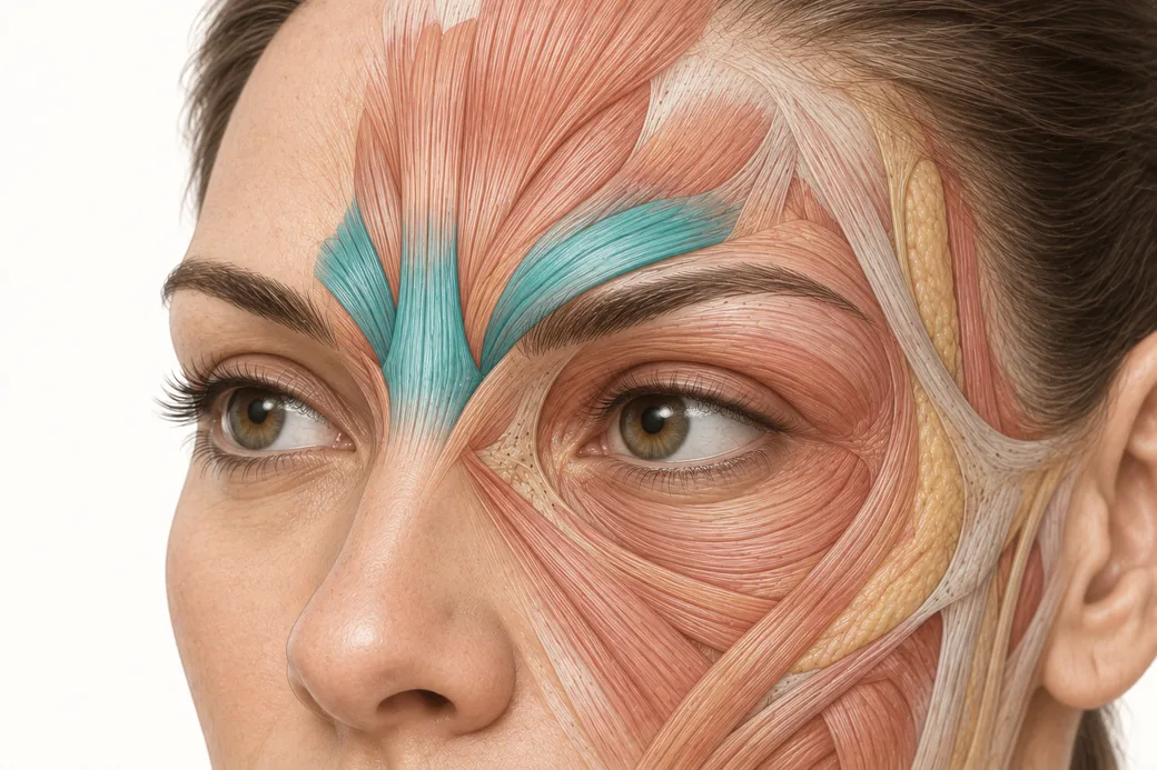
It meets the dorsal nasal artery, which is a branch of the ophthalmic artery. It sits immediately adjacent to the supratrochlear and supraorbital arteries, which are also ophthalmic artery branches. The ophthalmic artery is a branch of the internal carotid.
So at the medial canthus, the external carotid system and the internal carotid system shake hands. A filler embolus that enters the angular artery and is driven retrograde has an anatomical route into the ophthalmic artery and from there into the central retinal artery. That chain is the mechanism behind filler-induced vision loss, and it is worked through step by step in the companion spoke on the glabellar crossroad. (For the toxin-specific version of this question, which is a different mechanism entirely, see Can Botox cause blindness?.)
Two things about the medial canthus change how you should behave there. The vessel is shallow, so "superficial" is not protective. And the vessel is a junction, so an error there has more downstream destinations than an error anywhere else on the face.
The map has a second axis: depth
Naming vessels is where anatomy education usually stops. Knowing the name of something is not where the story ends.
The face is organised in five concentric layers — skin; subcutaneous fat; the superficial musculoaponeurotic system (SMAS) and its continuation into the mimetic muscles; the deep fat and areolar plane; and periosteum or deep fascia (Cotofana et al., JDDG, 2019). Every vessel on this map occupies a layer at every point along its course, and it does not stay in one layer.
That means "where is the facial artery?" is an incomplete question. The complete question is "where is the facial artery, at this point on the face, in which layer?" A needle placed at 3 mm is in a different anatomical world at the mandibular border than it is at the medial canthus, even though the number on the ruler is identical.
The layer axis is developed in its own spoke in this cluster. For the purposes of the map, hold one rule: depth is a coordinate, not a technique.
The map is probabilistic, not deterministic
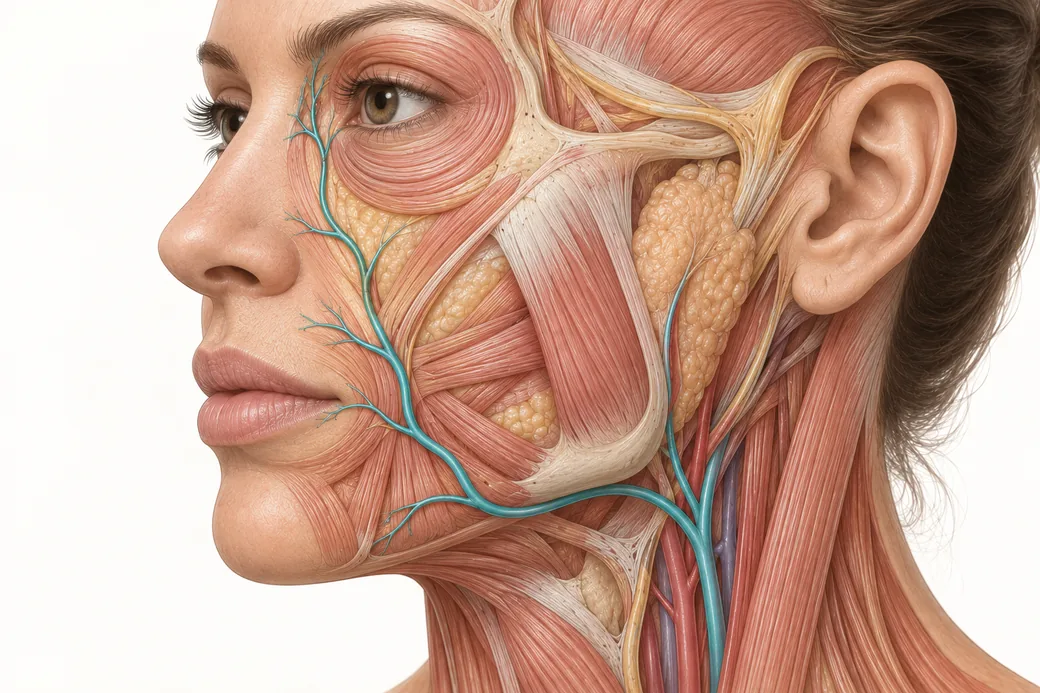
The facial artery has more anatomical variability than almost any vessel you will work around. It can be markedly tortuous. It changes depth along its course. It can terminate early. It can be duplicated. It can be hypoplastic.
The numbers are not subtle. In the 102-hemiface series, eight different termination points and 35 distinct branching-pattern combinations were identified. The angular artery — the terminus most injectors picture as the default — was present in only 33.33% of hemifaces in that study, and the reported prevalence across the wider literature ranges from roughly 32% to 74%. Yang and colleagues found the facial artery crossed the nasolabial fold in 33.9% of cases and ascended within 5 mm of it in 42.9% (Plastic and Reconstructive Surgery, 2014;133(5):1077–1082).
Read those figures the right way. They do not say the map is useless. They say the map gives you probabilities, not certainties, and that the correct response to a probability is a margin of safety, not a false sense of precision. A landmark tells you where a vessel is likely to travel. It does not tell you where this patient's vessel is.
This is the single best argument for cadaver work, and it is why I love teaching the anatomical cadaver class. You can read that a vessel is variable. It lands differently when you open ten faces and find ten different arteries.
The map is a network, not a tree
The last property of this map is the one that reframes everything.
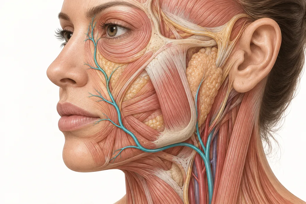
The branches of the facial artery are not endpoints. They anastomose — they form direct connections — with each other and with vessels from other territories. The superior labial connects across the midline and up into the nose through the columellar branch. The angular artery connects with the dorsal nasal artery. The lateral nasal artery connects into the nasal vascular arcade. The supratrochlear artery has documented anastomoses with the angular, supraorbital, dorsal nasal and superficial temporal arteries (Merizaj et al., Cureus, 2025;17(9):e92563).
The clinical consequence is the one most injectors are least prepared for: the complication frequently does not appear where you injected. You treat a lip and the patient calls about skin changes on the nose, or the forehead. That is not a mystery. That is the network behaving exactly as it is built.
When something goes wrong, the question is not only "where did I inject?" It is "where is this territory connected to, and where upstream is the obstruction most likely sitting?" Those are different questions and they produce different treatment maps. The dedicated spoke on anastomoses works through how that reframes a post-procedure phone call.
What this changes at the chairside
Four things, and none of them are new techniques. They are changes in what you do with the thirty seconds before you inject.
Survey before you plan. Stop. Look at the face and trace the route — notch, ascent, commissure, ala, canthus — before you choose a single entry point. You are asking not "why am I injecting here?" but "what is underneath here, and what does it connect to?"
Treat depth as part of the plan, not an afterthought. Decide the plane deliberately and know what that plane contains in that region. The relevant layer changes as you move up the face even when your needle length does not.
Adapt the whole approach, not just the site. Product choice, plane, needle versus cannula, injection speed and volume are all safety variables. Aspirating and injecting slowly matter. So does choosing a rheology and a tool appropriate to the territory.
Build the margin where the map is least certain. In regions with the highest anastomotic density and the shallowest terminal vessels — the nose, the glabella, the midface — probability thinking should make you more conservative, not more confident.
We have emergency protocols for vascular occlusion, and they are essential. But protocols are reactive by definition: a complication occurred and you are responding to it. Anatomy is proactive. Knowing the map lowers the number of times you have to open the protocol at all.
This map also pairs with a second one. Empire's faculty teach the trigeminal sensory map alongside the vascular map for a reason — the branch points, foramina and territories of V1, V2 and V3 sit in the same neighbourhoods as these arteries, and an injector who carries both maps is reasoning in three dimensions rather than two. Dr. Jennifer Thomas-Goering's trigeminal nerve material is the companion piece to this one.
Where to learn a map with your hands
You can memorise this page. I would rather you dissect it.
Empire Medical Training's Special Anatomical Cadaver Aesthetics Training and Anatomical Based Aesthetics Training are built around exactly this problem: seeing real vessels, at real depths, with real variability, in real tissue. For injectors applying the map to filler placement specifically, Complete Dermal Filler Training and Master Eye and Nose Injection Training work the regions where this map is least forgiving.
These anatomical relationships reflect Melissa Pulcini-Buttine's teaching in Empire Medical Training's hands-on curriculum, supported by the published anatomical literature cited above. Technique is learned under supervision; this article is educational and is not a substitute for training.
Melissa Pulcini-Buttine, PA — physician assistant of two decades; professor of anatomy and physiology for ~14 years; faculty member, Empire Medical Training; founder of an aesthetics practice in Greenwich, Connecticut.
Every guide in this cluster
 Clinical GuideAnastomoses: Why the Complication Doesn't Appear Where You InjectedFacial artery anastomoses explain why a lip treatment causes nasal skin changes. How arterial connections reframe injector triage and p
 Clinical GuideFiller Vascular Occlusion and Vision Loss: The Glabellar Crossroad, Step by StepHow filler vascular occlusion at the glabella causes vision loss — the retrograde embolic chain from the angular artery to the retina,
 Clinical GuideFacial Artery Variability: Why Landmarks Are Probabilities, Not CertaintiesFacial artery variability in cadaver data — termination points, branch prevalence, tortuosity and depth, and what probabilistic anatomy
 Clinical GuideThinking in Layers: Depth as the Second Axis of the Vascular MapFacial layers explained for injectors — the five-layer model, which layer each facial artery occupies, and why a depth in millimetres m
 Clinical GuideThe Perioral–Nasal Corridor: Labial, Columellar and Alar Arterial AnatomyLabial artery anatomy for injectors — superior and inferior labial arteries, the columellar branch and the arterial corridor linking th
 Clinical GuideGlabella Filler Risk: The Anatomy That Makes This the Least Forgiving TerritoryWhy glabella filler risk is the highest on the face: supratrochlear and supraorbital anatomy, internal carotid anastomoses, and the pat
 Clinical GuideAspirate, Low Pressure, Small Volumes, Stop Early: Four Habits That Reduce Vascular ComplicationsAspirate, low pressure, small volumes, stop early. What the evidence actually supports for each habit — including the genuine dispute o
 Clinical GuideNeedle vs Cannula Filler Decisions in High-Risk Regions: Neither Is 100% SafeNeedle vs cannula for filler in high-risk facial regions. What the occlusion data, arterial penetration studies and blindness registrie
 Clinical GuideRetrograde Embolisation: How Injection Pressure Sends Filler Where You Didn't Put ItHow retrograde embolisation works: the injection pressures involved, the volumes that matter, how filler fragments, and why product tra
Train with Empire
This guide is clinical education. The technique behind it is taught hands-on, on live patients, with faculty beside you.
Explore Anatomical-Based Aesthetics Training →Disclaimer
This article reflects the clinical opinions and experience of Melissa Pulcini-Buttine, PA, an independent faculty member contributing to Empire Medical Training's curriculum. The views expressed are the author's own and do not necessarily represent those of Empire Medical Training.
It is professional education, not medical advice, and is no substitute for hands-on training or independent clinical judgment. Licensed clinicians remain responsible for their own patient selection, technique and outcomes, for verifying current product labelling, and for practising within their scope and applicable law. Empire Medical Training accepts no liability for reliance on this content.
 Claim Your Prize!