








Filler vascular occlusion severe enough to threaten sight is not a mystery event. It is a mechanical sequence with identifiable steps, and every step in it is anatomical. If you can recite the chain, you can see where your own practice interrupts it — and where it doesn't.
The chain runs from a needle tip in the glabella or the upper midface, into an artery, backwards against the direction of blood flow, into a circulation that belongs to the internal carotid rather than the face, and finally into the vessels that supply the retina. It takes less material than most injectors assume and less time than anyone would like.
This page walks that chain step by step. It is deliberately anatomical rather than procedural: the recognition criteria and the emergency response belong in Empire's vascular occlusion material, and the separate question of botulinum toxin and vision is answered in a companion article on whether botulinum toxin can cause blindness — a different agent, a different mechanism, and not what this page is about.
Why the glabella is a crossroad, not just a wrinkle
The glabella is a small territory. That is the first problem. It is a few square centimetres of skin over a shallow bony platform, with very little soft tissue to absorb an error.
The second problem is that it is where four arterial systems meet.
Ascending from below is the angular artery — the terminal segment of the facial artery, which arrives at the medial canthus and, on ultrasound, sits roughly 1 mm from the skin surface there. The facial artery belongs to the external carotid system.
Descending from above are the supratrochlear and supraorbital arteries, which exit the orbit at the superior orbital rim. Alongside them is the dorsal nasal artery, running onto the nasal dorsum. All three are branches of the ophthalmic artery, which is a branch of the internal carotid.
And these systems anastomose. The supratrochlear artery has documented, consistent connections with the angular, supraorbital, dorsal nasal and superficial temporal arteries (Merizaj et al., Cureus, 2025;17(9):e92563). That is the crossroad. Within a couple of centimetres, the vascular supply of the face and the vascular supply of the eye become one continuous lumen.
The third problem is depth. These are not buried vessels. The supratrochlear artery exits the orbit approximately 15–18 mm from the facial midline and runs subcutaneously for much of its course — its superior third beneath the dermis and above the fat, its inferior two-thirds above the frontalis and beneath the fat. Ultrasound of the central forehead measured the superficial branch at 4.2 mm depth with a 14.7 mm lateral offset, and the deep branch at 5.9 mm and 19.2 mm (Phumyoo et al., Clinical Anatomy, 2020). Cadaveric work cited in the same literature identifies high-risk planes in the glabella and medial forehead at roughly 4.1–5.1 mm.
A small territory, a dense convergence of superficial vessels, and a direct lumen to the eye. That is what "high-risk region" means anatomically. It is not a label on a diagram; it is a description of what is underneath.
The chain, step by step
Step 1 — The needle tip enters a lumen
Nothing in the chain happens without this. An intravascular needle or cannula tip is the necessary first condition, and it is silent. There is no reliable tactile signal. Aspiration is an imperfect check: in one in vitro assessment of seventeen filler products, aspiration was positive in only 53% (Casabona, Dermatologic Surgery, 2015), and the current consensus position in the aesthetic literature is that neither a positive nor a negative aspiration should be relied upon as a safety manoeuvre (van Loghem et al., Aesthetic Surgery Journal, 2022;42(1):89–101).
That is not an argument against aspirating. It is an argument against treating aspiration as the thing standing between your patient and an occlusion.
Step 2 — Injection pressure exceeds the arterial pressure ahead of it
For filler to move backwards in an artery, the pressure at the needle tip has to exceed the sum of systolic arterial pressure and the frictional resistance of the column of blood and product it is pushing against. A thumb on a plunger can generate that. This is the single most important reason that injection speed and force are safety variables and not stylistic preferences.
Below that threshold, material entering a lumen is carried distally with flow. Above it, the column is driven proximally — toward the origin of the vessel and toward the junctions where the territories meet.
Step 3 — The retrograde column forms and travels toward the orbit
Once flow is reversed locally, the material fills the vessel backwards. A needle tip in the glabella that is inside the supratrochlear artery is, in that moment, a short distance from the orbital apex.
How short is the most sobering number in this literature. Cadaveric dissection of six fresh tissue heads (twelve hemifaces) measured the entire volume of the supratrochlear artery from the glabella to the orbital apex at a mean of 0.085 mL (Khan et al., Aesthetic Surgery Journal, 2017;37(2):203–208).
Eighty-five thousandths of a millilitre. Less than a tenth of a syringe graduation. That is the volume that fills the pipe from a glabellar injection point to the junction with the ophthalmic artery.
Step 4 — Pressure is released and the material redistributes
When the plunger stops, the driving pressure disappears and normal arterial pressure reasserts itself. The column of material — now sitting proximally, at or near the ophthalmic artery — is then pushed by native flow into whatever branch geometry offers.
This is why the injury is not confined to the vessel entered, and why the resulting picture is often mixed: some material travels onward into the eye's arterial supply, some is redistributed into cutaneous branches, producing skin ischaemia in the same event.
Step 5 — Occlusion in the ocular circulation
The ophthalmic artery gives rise to the central retinal artery and the posterior ciliary arteries. Occlusion at this level produces retinal ischaemia. The clinical presentation is sudden and usually profound: abrupt vision loss, frequently with pain, and often accompanied by ocular motility changes or ptosis when adjacent orbital vessels are involved.
Step 6 — The ischaemic clock
Retinal tissue does not tolerate arterial occlusion for long, and the window in which intervention has any realistic prospect of altering outcome is measured in a small number of hours at most. This is why every serious discussion of this complication ends in the same place: prevention, and then recognition speed.
What the published case record actually shows
The case literature is now substantial enough to be informative rather than anecdotal, and it corrects several assumptions injectors carry.
The most comprehensive review to date identified 365 new cases of filler-associated vision loss published between September 2018 and March 2023, bringing the cumulative published total to 511 cases since 1906 (Doyon et al., Aesthetic Surgery Journal, 2024;44(10):1091–1104). An earlier update had identified 48 cases between 2015 and 2018, for a running total of 146 (Beleznay et al., Aesthetic Surgery Journal, 2019;39(6):662–674). The acceleration in reported cases is not subtle.
Three findings from that record deserve to change behaviour.
The site distribution is not what most injectors expect. In the 2018–2023 cases, the most common injection site was the nose at 40.6%, followed by the forehead at 27.7%, then the glabella at 19.0%. The glabella is the classically taught high-risk site, and it is third. The nasal and forehead territories — both sitting on the same crossroad through the dorsal nasal, lateral nasal and supratrochlear connections — together account for more than two-thirds.
Recovery is the exception. Among 318 cases reporting visual outcome, 68.2% had no recovery, 25.8% had partial improvement, and 6.0% recovered completely. This is not a complication that usually resolves.
It is rarely an isolated eye event. Skin changes were present in 73.2% of cases, and CNS involvement in 34.0%, with strokelike features in 19.2%. Hyaluronic acid accounted for 79.6% of cases in the recent cohort — which reflects usage volume, not a claim that other agents are safe.
That last cluster of figures has a direct clinical reading: ischaemic skin findings after an injectable in the midface, nose or upper face are not only a skin problem. They are a signal that material went intravascular, and vision must be assessed.
Why "small volume, slow, small syringe" is an anatomical argument
Injectors often hear these as generic caution. Read against the chain above, they are specific mechanical interventions at identified steps.
Slow injection attacks step 2. Pressure at the needle tip is what reverses flow. Lower the rate and you lower the peak pressure.
Small aliquots attack step 3. If the volume that can reach the orbital apex from a glabellar vessel is on the order of 0.085 mL, then any single uninterrupted bolus approaching that magnitude is a meaningful quantity in the wrong lumen.
Moving the tip, not injecting on a static point reduces the time any single lumen is exposed to sustained pressure.
Cannula selection in appropriate regions attacks step 1 — a blunt tip is less likely, though not unable, to enter a lumen. Melissa Pulcini-Buttine teaches needle-versus-cannula choice, injection speed, aspiration and volume as a package of adaptive decisions made after you have surveyed the vascular territory, not as a fixed house style. The point is that each of these levers maps onto a step in a mechanism, and knowing which step is what lets you reason about them rather than follow them.
None of these reduce risk to zero. In anatomy there is no 0% risk area, and there is no manoeuvre that makes one.
The three regions where this chain can start
From this transcript and this map, three regions carry the highest risk for the sight-threatening version of this chain: the nose, the glabella and the midface. They are high-risk for one shared reason — each sits on, or connects directly into, the arterial junctions that lead to the ophthalmic circulation.
The nose is supplied by the lateral nasal and dorsal nasal arteries, which anastomose with the angular artery and the ophthalmic system. The glabella hosts the supratrochlear and supraorbital arteries directly. The midface carries the angular segment of the facial artery toward the medial canthus, and published ultrasound work on the angular artery in the midface concludes explicitly that there is no guaranteed safe location in this region (Cotofana et al., Aesthetic Surgery Journal, 2021;41(7):805–813).
That last conclusion is worth sitting with. It is not a rhetorical caution. It is the measured finding of anatomical variability applied to a specific region.
What this changes at the chairside
Treat the nose as the highest-risk site on the face, not the glabella. The published case distribution says so. If your mental risk ranking still puts the glabella first, update it.
Assume proximity to a shallow vessel in the glabella, always. With the supratrochlear artery running subcutaneously for most of its course and high-risk planes reported around 4–5 mm, superficial placement is not inherently protective here. It may be the opposite.
Ask about vision whenever you see ischaemic skin. In 73.2% of published vision-loss cases there were skin changes. Cutaneous findings are the visible end of an event whose other end may be ocular.
Treat pressure and volume as the controllable variables. You cannot control anatomical variation. You can control how hard and how fast you push, and how much goes in before you stop and look.
Know your escalation route before you need it. Not the protocol details — those belong in Empire's vascular occlusion recognition and response material — but the practical answer to "who do I call and how fast can this patient be in front of an ophthalmologist?" That answer should exist before the day you need it, not be assembled on the day. On the pharmacology of dissolving hyaluronic acid specifically, see how filler is dissolved.
Learning the crossroad in tissue
This is the region where reading about anatomy and seeing anatomy diverge most sharply. The supratrochlear artery is a millimetre-scale vessel in a shallow plane whose position varies between individuals and between the two sides of the same face. A diagram cannot convey that. A dissection can.
Empire Medical Training's Special Anatomical Cadaver Aesthetics Training and Anatomical Based Aesthetics Training are structured around exactly this kind of region-specific vascular anatomy, and Master Eye and Nose Injection Training addresses the two territories the case record identifies as highest risk.
These anatomical relationships and mechanistic steps reflect Melissa Pulcini-Buttine's teaching in Empire Medical Training's hands-on curriculum, supported by the published literature cited above. This article is educational, is not a substitute for supervised training, and does not replace your practice's vascular complication protocol.
Melissa Pulcini-Buttine, PA — physician assistant of two decades; professor of anatomy and physiology for ~14 years; faculty member, Empire Medical Training; founder of an aesthetics practice in Greenwich, Connecticut.
Related guides in this cluster
Part of Facial Vascular Anatomy for Injectors.
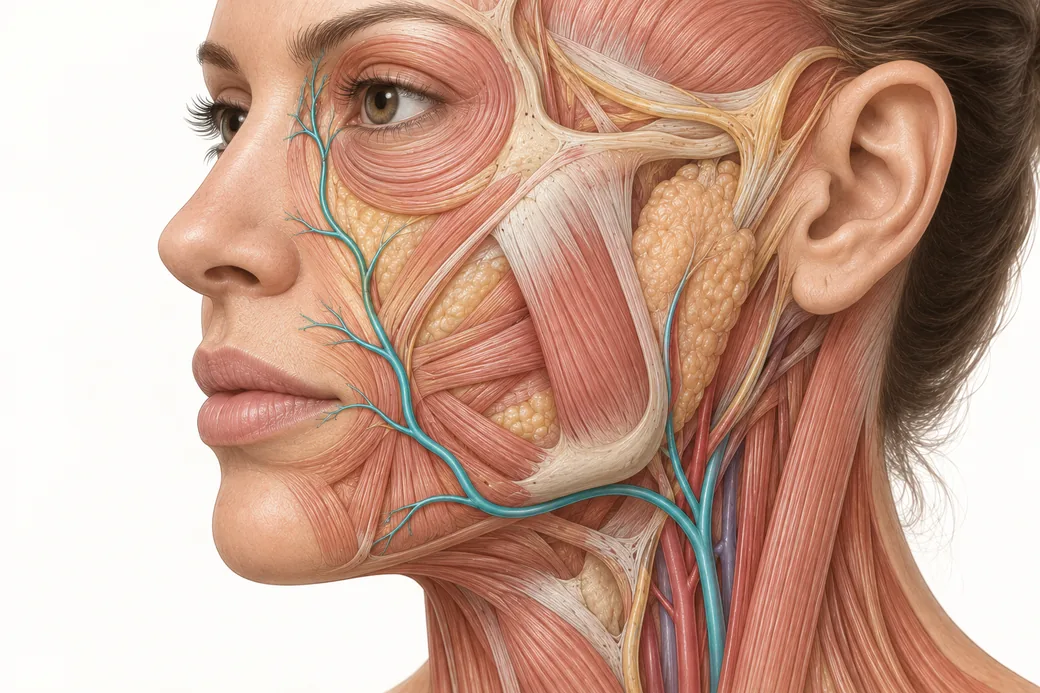 Clinical GuideAnastomoses: Why the Complication Doesn't Appear Where You InjectedFacial artery anastomoses explain why a lip treatment causes nasal skin changes. How arterial connections reframe injector triage and p
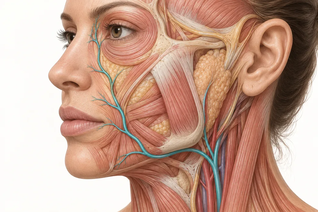 Clinical GuideFacial Artery Variability: Why Landmarks Are Probabilities, Not CertaintiesFacial artery variability in cadaver data — termination points, branch prevalence, tortuosity and depth, and what probabilistic anatomy
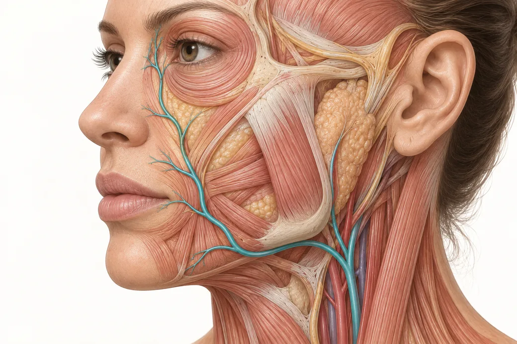 Clinical GuideThinking in Layers: Depth as the Second Axis of the Vascular MapFacial layers explained for injectors — the five-layer model, which layer each facial artery occupies, and why a depth in millimetres m
Train with Empire
This guide is clinical education. The technique behind it is taught hands-on, on live patients, with faculty beside you.
Explore Anatomical-Based Aesthetics Training →Disclaimer
This article reflects the clinical opinions and experience of Melissa Pulcini-Buttine, PA, an independent faculty member contributing to Empire Medical Training's curriculum. The views expressed are the author's own and do not necessarily represent those of Empire Medical Training.
It is professional education, not medical advice, and is no substitute for hands-on training or independent clinical judgment. Licensed clinicians remain responsible for their own patient selection, technique and outcomes, for verifying current product labelling, and for practising within their scope and applicable law. Empire Medical Training accepts no liability for reliance on this content.
 Claim Your Prize!