








Glabella filler risk is not a matter of opinion or of injector confidence. It is a matter of which carotid system you are standing in. The glabella is a small territory — a few square centimetres of skin between the brows — and almost every injector underestimates it for exactly that reason. It looks like a simple furrow. It is, anatomically, the one place on the anterior face where the internal carotid circulation surfaces into injectable soft tissue, and that single fact reorganises everything about how the region should be treated.
"It's a very small territory, but there's a lot around there," is how Melissa Pulcini-Buttine, PA, frames it when she teaches the region. "Low margin for error." That phrase is the whole piece. Margin for error is not a function of how steady your hand is. It is a function of how much anatomical slack exists between where your needle tip is and where a catastrophic outcome begins. In the glabella, that slack is measured in millimetres, and the consequence at the end of it is not a bruise or a nodule. It is the retina.
This article is a regional risk reference for the glabellar complex. It sits inside Empire's facial vascular anatomy cluster; the full-face arterial map and the territory-by-territory course anatomy live at the cluster pillar. What follows is specifically about why this region behaves differently from the rest of the face, and what that should change about your hands.
The carotid problem: most of the face is not like the glabella
Work out from the midline and the face is overwhelmingly external carotid territory. The facial artery, the superficial temporal artery, the infraorbital and the transverse facial artery are all external carotid derivatives. An embolic event in pure external carotid territory is a serious complication — skin necrosis, scarring, a patient who will remember you for the rest of their life — but it is a soft tissue complication.
The glabella is different. The supratrochlear artery and the supraorbital artery are terminal branches of the ophthalmic artery, and the ophthalmic artery is the first major intracranial branch of the internal carotid artery. The dorsal nasal artery, emerging at the medial canthus, belongs to the same system.
This is the crux of glabellar risk and it is worth stating without hedging: when you inject the glabella, you are injecting into vessels that drain, retrogradely, toward the internal carotid circulation. The central retinal artery is a branch of the ophthalmic artery. There is a continuous, patent, anatomically unavoidable channel between the tip of your needle in the glabellar furrow and the blood supply of the eye.
Melissa puts it plainly in the cadaver lab: those vessels "can anastomose with the ophthalmic artery, get to the retinal artery, and then you can literally have permanent blindness."
That is not a rare-event footnote appended for medico-legal completeness. It is the defining anatomical feature of the territory, and it is why the glabella should be reasoned about differently from the cheek, the jawline or the temple — not because the injection is harder, but because the failure mode is categorically worse.
The supratrochlear artery and why "find the safe plane" fails here
Injectors are trained, correctly, to think in planes. Supraperiosteal here, deep dermal there, subcutaneous in between. The implicit promise of plane-based technique is that there exists a depth at which you are not in a vessel.
The glabella does not honour that promise, and the reason is that the supratrochlear artery changes depth as it ascends.
At the level of the superomedial orbital rim the vessel is deep — running beneath the corrugator supercilii, close to bone, in the company of the supratrochlear nerve. As it climbs the central forehead it pierces the muscle and becomes progressively more superficial, ultimately running in the subcutaneous plane. The transition is not a sharp line and it is not identical between two patients.
The practical consequence is the part that matters at the chairside: there is no single plane through the glabella and lower central forehead that is reliably avascular. Go deep at the rim and you are in the neurovascular bundle. Go superficial on the forehead and you are in the same vessel further along its course. The plane that is safe at one point on the vessel is the plane that is dangerous two centimetres away.
And the height at which that transition happens is not predictable in an individual patient. An ultrasound investigation of 50 living subjects measured where the supratrochlear and supraorbital arteries change plane relative to the superior orbital rim and found a mean of roughly 13 to 14 mm — with a range of 4.0 to 27.0 mm (Cotofana S, Velthuis PJ, Alfertshofer M, et al. The Change of Plane of the Supratrochlear and Supraorbital Arteries in the Forehead — An Ultrasound-Based Investigation. Aesthet Surg J. 2021;41(11):NP1589-NP1598. PMID 33652475). The authors concluded that the superficial lower forehead "should be avoided due to the unpredictability and inconsistent presence of the central and paracentral arteries."
A mean of 14 mm with a range of 4 to 27 mm is not a landmark. It is a warning that the number you would want to act on does not exist for the patient in your chair.
This is why the glabella defeats the injector whose safety model is entirely procedural. A procedural model says "deep here, superficial there" and stops. An anatomical model asks where this particular vessel is in this particular patient at this particular point, and accepts that the answer is probabilistic rather than certain.
Anatomical variability is not a caveat you add at the end. In the glabella it is the operating condition. The supratrochlear artery has a described course, not a fixed one, and the distance between its described course and where it actually is in the patient in front of you is the entire margin you are working with.
The supraorbital foramen: the landmark you can actually feel
Almost nothing in facial vascular anatomy can be confirmed by touch. The supraorbital neurovascular bundle is the exception, and Melissa treats it as a non-negotiable step rather than an optional nicety.
Run your thumb along the superior orbital rim and you will find the supraorbital notch — in a proportion of patients a true closed foramen rather than an open notch. The supraorbital artery and the supraorbital nerve exit together at that point. It is palpable, it is patient-specific, and it takes four seconds.
"Touch your patients, feel them, mark them, have them move," she teaches. "Then you could really assess where these things are."
Three things follow from palpating it:
- You have localised a real vessel in a real patient rather than a vessel in a textbook. Whatever the atlas says about the average position of the supraorbital bundle relative to the midpupillary line, you have just measured this one.
- You have established a reference point for your own markings. Everything else in the region can be marked relative to a landmark you have physically confirmed.
- You have identified a foramen you do not want to inject into or toward. This matters for filler and, separately, for toxin — the foramen is a route by which product placed at the rim can travel where you did not intend it to. That toxin-specific pathway, and the true eyelid ptosis it can produce, is covered in the neurotoxin anatomy cluster.
Palpation does not make the glabella safe. Nothing makes the glabella safe. It converts one unknown into a known, which in a region with this little margin is a meaningful trade.
Anastomoses: why external carotid injections reach internal carotid territory
If the glabellar vessels were an isolated island of internal carotid anatomy, the risk would be confined to glabellar injections. They are not.
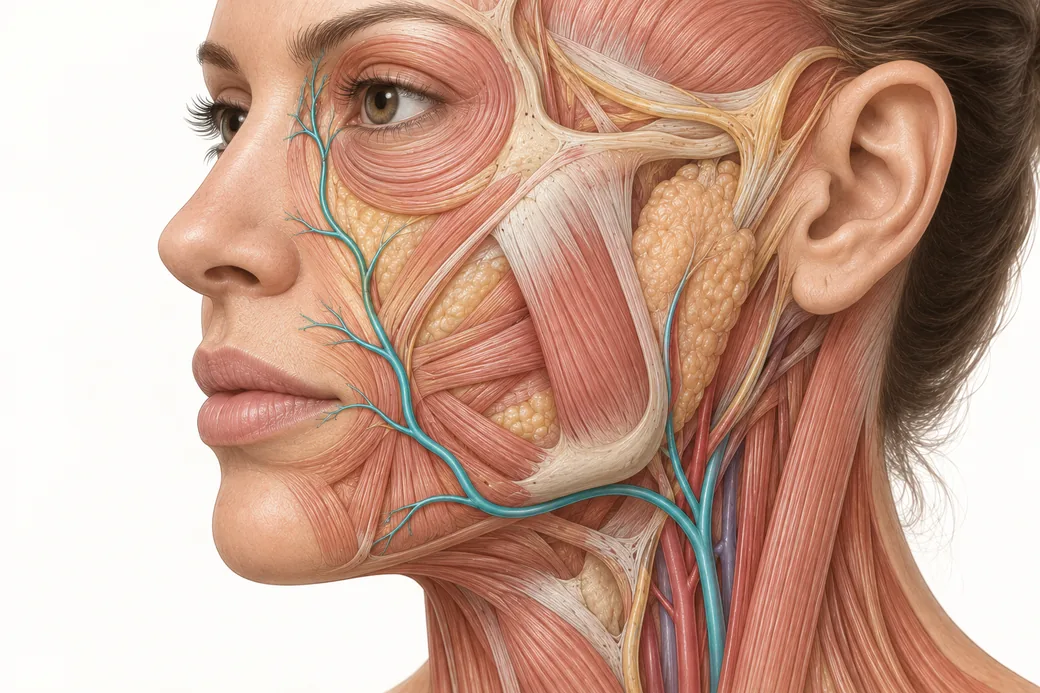
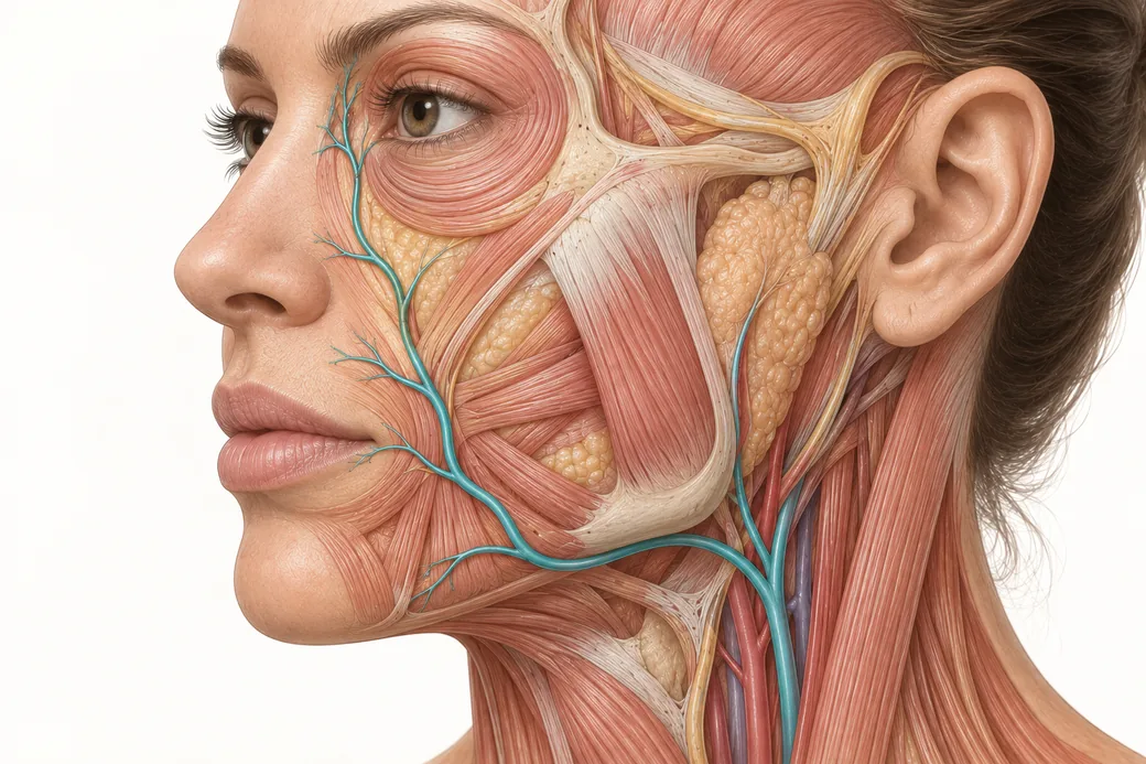
The angular artery, the terminal branch of the facial artery, ascends alongside the nose to the medial canthus and anastomoses with the dorsal nasal artery — an ophthalmic artery branch. The supratrochlear and supraorbital arteries anastomose with the superficial temporal system laterally and across the midline with their contralateral partners. The face is not a set of separate vascular territories. It is a connected network with watershed zones, and the medial canthus is one of the most important of them.
Melissa returns to this word repeatedly — anastomosis, "the communication between other vessels" — because it is what makes the region's risk non-local. A bolus placed in the nasal dorsum, the medial nasolabial fold or the medial canthal region is not necessarily confined to external carotid territory. Under sufficient injection pressure it can be driven retrogradely into a communicating channel and arrive somewhere you never touched.
This is the anatomical basis for something that confuses injectors when it happens: ischaemic change appearing in skin you did not inject. Blanching in a territory distant from the needle is not a mystery. It is the anastomotic network doing exactly what it is built to do, with your product in it.
The hemodynamics of how that transfer actually occurs — the pressure relationships that let a syringe overcome arterial flow — are the subject of a separate spoke in this cluster. What matters regionally is this: the glabella's risk radius is larger than the glabella.
Why toxin and filler are not the same conversation here
Melissa is explicit that neuromodulator and filler are different treatments doing different things, and that lumping them into a single "glabellar danger" warning muddles the teaching. "Botox is going to affect muscle movement. Filler is going to replace volume. So they do two different things."
They also fail differently.
- Toxin complications in the glabella are complications of diffusion and muscle selection. Product reaches a muscle you did not target — the frontalis, the levator palpebrae superioris — and you get brow ptosis or true eyelid ptosis. Distressing, sometimes weeks of it, but self-limiting.
- Filler complications in the glabella are complications of vascular occlusion. Product reaches a vessel you did not intend, and the result is ischaemia — potentially skin necrosis, potentially visual loss. Not self-limiting.
Both are worth teaching. They are not the same order of risk, and conflating them produces injectors who are appropriately anxious about the wrong thing. If you want the toxin-side companion to this article, the question of whether neuromodulator can produce visual loss is handled separately in can Botox cause blindness.
Melissa's own sequencing preference follows from the distinction. "I usually love to start with toxin no matter what, before doing any filler," she teaches — relax the dynamic component first, reassess what volume deficit genuinely remains, and in a number of patients discover that the glabellar furrow softened enough that the filler conversation changes. That is her practice pattern, not a universal standard, but the logic behind it is sound: the safest glabellar filler injection is the one the patient turned out not to need.
What "low margin for error" actually asks of you
Melissa's guidance for the region reduces to a short and unglamorous list, and she teaches it as a sequence rather than a menu.
Know the structures before the needle moves. Not "know that there are vessels there" — know which vessels, from which carotid system, at which depth, and what they communicate with. In Empire's cadaver dissections the point of opening the glabellar complex is precisely that you cannot build this from a diagram. You build it from having seen the supratrochlear bundle emerge and watched where it goes.
Palpate and mark, with the patient animating. Static assessment misses the muscle borders. Have them make an angry face, mark what actually moves, find the supraorbital notch, and work from your own marks rather than from remembered proportions.
Aspirate, and believe a positive aspiration. "If you have a positive aspiration, do not push any product," Melissa teaches, "because then you can spread it to the vessels that anastomose to something dangerous." The evidence around aspiration is genuinely contested and is examined honestly in a separate spoke of this cluster — but the asymmetry is worth noting: a positive aspiration is highly informative, even though a negative one proves much less than injectors assume.
Low pressure, slow delivery, small volumes. "Injection pressure overcomes the local flow. Press slow." A high-pressure bolus is the mechanism by which the anastomotic network gets loaded.
Stop early. Melissa's phrasing is "stop early, because it can escalate fast." The glabella is not a region in which to push through an unexpected finding — unusual pain, blanching, resistance that does not feel right — and reassess afterwards. Reassess first.
Have the rescue protocol ready before you need it. Hyaluronidase on the shelf, dosing decided, referral pathway known. Assembling a response during an event is how minutes are lost, and in ischaemia minutes are the currency. What dissolution can and cannot achieve is covered in filler dissolve.
The honest summary
The glabella is not dangerous because it is technically difficult. Technically it is one of the simpler injections on the face. It is dangerous because it is the one anterior facial region where a small error has access to the internal carotid circulation, because the vessel changes plane as it ascends so no single depth is reliably safe, and because the anastomotic network means the consequence need not appear where the needle was.
"Respect anatomy," as Melissa puts it. "Knowledge is power." That sounds like a slogan until you notice that in this territory it is a literal description of the only protective factor available. You cannot see the vessel. You cannot reliably feel it. You cannot avoid it with a plane. What you can do is know it is there, know where it goes, and inject as though it is exactly where you would least like it to be.
These figures and techniques reflect Melissa Pulcini-Buttine's clinical practice as taught in Empire Medical Training's hands-on curriculum. Technique is learned under supervision; this article is educational and is not a substitute for training.
Injectors who want to build this from dissection rather than diagrams should look at Empire's anatomical based aesthetics training and the special anatomical cadaver aesthetics training, where the glabellar complex is opened and traced. Injectors building core filler competence will find the regional foundations in complete dermal filler training, and those working in the highest-risk periorbital and nasal territories in master eye and nose injection training.
Melissa Pulcini-Buttine, PA — physician assistant of two decades; professor of anatomy and physiology for ~14 years; faculty member, Empire Medical Training; founder of an aesthetics practice in Greenwich, Connecticut.
Related guides in this cluster
Part of Facial Vascular Anatomy for Injectors.
 Clinical GuideAnastomoses: Why the Complication Doesn't Appear Where You InjectedFacial artery anastomoses explain why a lip treatment causes nasal skin changes. How arterial connections reframe injector triage and p
 Clinical GuideFiller Vascular Occlusion and Vision Loss: The Glabellar Crossroad, Step by StepHow filler vascular occlusion at the glabella causes vision loss — the retrograde embolic chain from the angular artery to the retina,
 Clinical GuideFacial Artery Variability: Why Landmarks Are Probabilities, Not CertaintiesFacial artery variability in cadaver data — termination points, branch prevalence, tortuosity and depth, and what probabilistic anatomy
Train with Empire
This guide is clinical education. The technique behind it is taught hands-on, on live patients, with faculty beside you.
Explore Anatomical-Based Aesthetics Training →Disclaimer
This article reflects the clinical opinions and experience of Melissa Pulcini-Buttine, PA, an independent faculty member contributing to Empire Medical Training's curriculum. The views expressed are the author's own and do not necessarily represent those of Empire Medical Training.
It is professional education, not medical advice, and is no substitute for hands-on training or independent clinical judgment. Licensed clinicians remain responsible for their own patient selection, technique and outcomes, for verifying current product labelling, and for practising within their scope and applicable law. Empire Medical Training accepts no liability for reliance on this content.
 Claim Your Prize!