







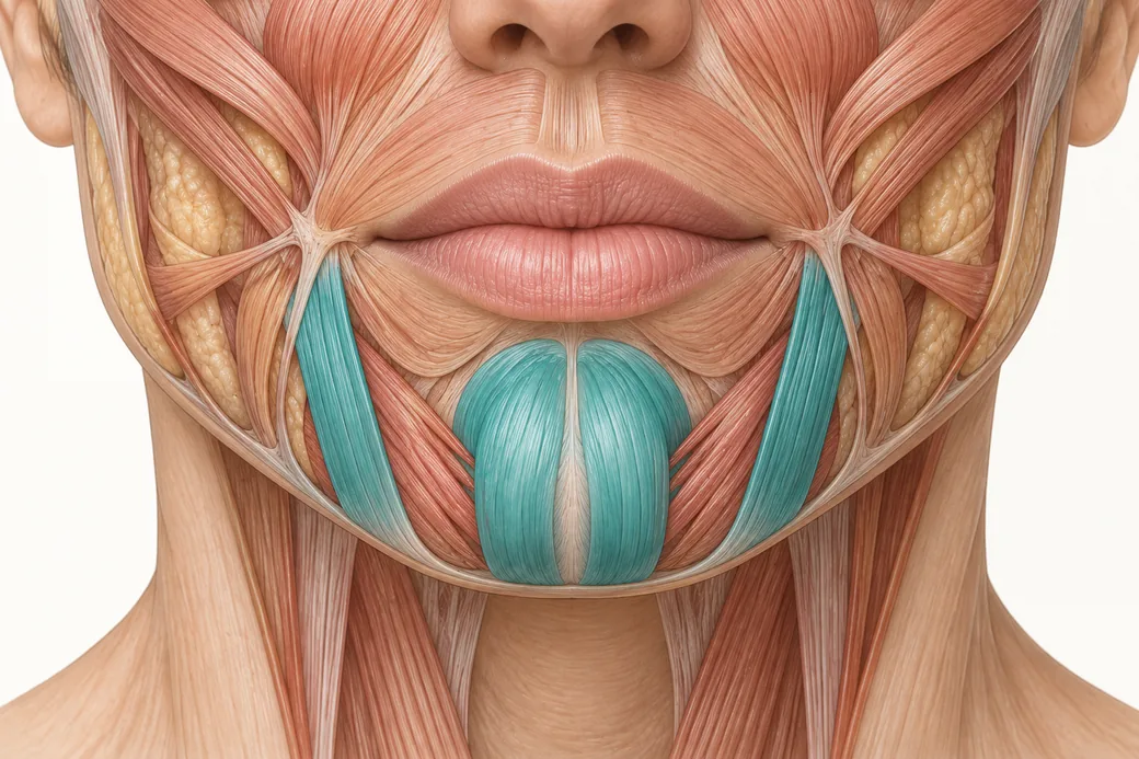
Labial artery anatomy is usually taught as a lip topic. It is not. The labial arteries are the middle section of a continuous arterial corridor that runs from the facial artery at the oral commissure, through both lips, up the philtrum into the columella, and onward into the nasal base and tip.
That corridor is the anatomical substrate for the single most confusing complication pattern in aesthetic practice: a patient treated in the lips who develops ischaemic skin changes in the nose. Once you can see the corridor as one unit rather than as a lip territory and a separate nasal territory, that pattern stops being a surprise and becomes a prediction.
This page is the anatomy of that corridor. It contains no lip injection technique, no product guidance and no placement recommendations — those are clinical decisions learned under supervision. What patients typically report after lip treatment, and how to separate expected findings from concerning ones, is covered separately in common lip filler reactions and how to avoid them.
What "corridor" means here
Most vascular diagrams draw the perioral region as a set of separate branches hanging off the facial artery: inferior labial to the lower lip, superior labial to the upper lip, a nasal branch somewhere above. Distinct arrows, distinct territories.
Anatomically that is misleading in two ways.
First, the labial arteries anastomose across the midline with their contralateral counterparts, forming a perioral arterial ring around the mouth. The lips are not two independently supplied halves; they are a loop with inflow from both sides.
Second, the superior labial artery does not stop at the philtrum. It commonly gives a branch that ascends into the columella and the nasal base. So the loop has an exit ramp, and the exit ramp goes to the nose.
A corridor, not a set of endpoints. That single structural fact is what the rest of this page describes in detail.
The inferior labial artery
The inferior labial artery arises from the facial artery near the oral commissure and runs medially through the lower lip toward the midline, where it anastomoses with its counterpart.
It is present in the large majority of dissections but not universally: 78.43% of 102 hemifaces in a recent cadaveric series (Nguyen et al., Archives of Craniofacial Surgery, 2024;25(2):77–84). Reported prevalence across the wider literature ranges from roughly 57.5% to 100%, which is a range wide enough to tell you the vessel is genuinely inconstant rather than merely difficult to dissect.
Ultrasound in 41 living volunteers put its mean depth in the lower lip at 5.2 ± 0.14 mm, and found it within the red lip rather than below the vermilion border in 86.2% of lower lips (Cotofana et al., Aesthetic Surgery Journal, 2020;40(12):1327–1335).
The superior labial artery
The superior labial artery is the more consistently present of the pair — 87.25% in the same 102-hemiface series, with a wider literature range of roughly 77.5% to 98.0%. It arises from the facial artery above the commissure and runs medially through the upper lip.
Three properties matter to an injector.
Depth. Mean 5.6 ± 0.13 mm in the upper lip on ultrasound.
Plane. This is the more useful measurement. Across the labial arteries, the most frequent location was the submucosal plane at 58.5%, followed by intramuscular at 36.2%, and subcutaneous at only 5.3%. The vessel is predominantly deep to or within the orbicularis oris relative to the skin surface, and only rarely in the subcutaneous layer.
Position relative to the vermilion. In 83% of upper lips the artery was found within the red lip rather than above the vermilion border. The vessel sits toward the wet side, not along the white roll.
A four-type classification of the superior labial artery describes the arrangements it forms with the alar branch: the facial artery independently giving rise to both (56.7%), the alar branch arising from the superior labial artery (21.7%), the facial artery terminating as the superior labial artery (15.0%), and the superior labial artery absent (6.7%).
Note the third of those. In roughly one in seven specimens, the facial artery ends as the superior labial artery — it never reaches the nose at all. In those patients the nasal supply arrives entirely from elsewhere, most often descending from the ophthalmic system. The corridor still exists; it is just being fed from the other end.
The columellar branch: the road from the lip to the nose
This is the branch that explains the phone call.
As the superior labial artery approaches the philtrum, it commonly gives off a columellar branch that ascends the philtral column to the nasal septum and base, entering the columella at the columellolabial junction.
The published prevalence figures are worth knowing precisely, because they are both high enough to matter and variable enough to prevent assumptions:
- In one series, the superior labial artery supplied the columellar branches in 72.5% of specimens (37 of 51), with the inferior alar ramus supplying them in the remaining 27.5%.
- In another, the columellar branch was visualised bilaterally in 9%, unilaterally in 68%, and was absent in 23% of specimens (Jung et al., The Laryngoscope, 2000).
Put those together and the working model is: a direct arterial road from the upper lip to the nasal base exists in most people, usually on one side only, and occasionally not at all.
The clinical corollary is the one Melissa Pulcini-Buttine teaches directly — you could be injecting the lip and the philtral column and affect the nose. If something is going on with the nose after a perioral treatment, the answer to "but I didn't inject the nose" is that you injected a vessel that goes there.
The septal branches and the nasal tip
Beyond the columella, the corridor continues into the nasal tip.
The deep septal branch of the superior labial artery arises from its middle portion, courses anteriorly just below the septal cartilage, emerges between the superior border of the orbicularis oris and the septal cartilage, and ascends toward the nasal tip in 90.0% of cases (Nguyen et al., Surgical and Radiologic Anatomy, 2025; doi:10.1007/s00276-025-03659-z). A superficial septal branch runs a more anterior course in the columellar soft tissue.
At the tip, these ascending branches meet vessels coming the other way. The lateral nasal artery and the dorsal nasal artery pass across the dome region and anastomose with the columellar arteries. That junction — at the nasal tip, between vessels ascending from the lip and vessels descending from the nasal sidewall and dorsum — is the upper end of the corridor and the point where it joins the facial-to-ophthalmic network described in this cluster's anastomoses spoke.
So the full route, bottom to top, is: facial artery → superior labial artery → columellar branch → septal branches → nasal tip → lateral nasal and dorsal nasal arteries → the angular and ophthalmic systems.
That is one continuous path from the mandibular border to the orbit, and every segment of it is a named vessel with published prevalence data.
The alar and lateral nasal arteries
The corridor's upper-middle section is supplied by the branches serving the nasal ala and sidewall.
The lateral nasal artery arises from the facial artery as it passes alongside the ala and supplies the nasal sidewall, ala and dorsum. It is the single most common termination point of the facial artery — the vessel ended there in 32.35% of the 102 hemifaces studied.
The inferior alar artery is a smaller, more variable vessel supplying the alar base; the facial artery terminated there in 6.86% of hemifaces, and as noted above, the alar branch's relationship to the superior labial artery is itself one of the axes on which perioral arterial anatomy varies.
The ala is therefore a convergence point: supplied from below by the corridor, from the side by the lateral nasal artery, and from above by the dorsal nasal artery. Small territory, thin soft tissue envelope, multiple inflows, and — critically — direct continuity with the ophthalmic circulation.
How variable is the corridor?
Every segment of it. That is the honest summary, and the figures above make the point better than any generalisation:
| Structure | Reported presence |
|---|---|
| Superior labial artery | 87.25% (range ~77.5–98.0%) |
| Inferior labial artery | 78.43% (range ~57.5–100%) |
| Columellar branch | bilateral 9%, unilateral 68%, absent 23% |
| Columellar supply from superior labial artery | 72.5% (remainder from inferior alar ramus) |
| Deep septal branch ascending to tip | 90.0% |
| Facial artery terminating as lateral nasal artery | 32.35% |
| Facial artery terminating as a labial artery | ~9.8% combined |
Two readings follow, and only one of them is correct.
The incorrect reading: if the vessel is often absent, the region is often safer. It is not. Absence of a named vessel means the territory is supplied by a different arrangement — a contralateral vessel crossing the midline, an enlarged counterpart, a branch from a neighbouring territory. Perioral and nasal tissue is richly perfused in everyone. Variability redistributes the plumbing; it does not remove it.
The correct reading: you cannot know which arrangement is in front of you. The corridor exists in essentially every patient; its specific route differs. That is an argument for margin, not for confidence.
Reading the corridor as one territory
The reason to learn this as a corridor rather than as four separate vessels is that it changes what you examine and what you ask.
If the perioral region and the nasal base are one arterial territory, then a perioral treatment has a nasal territory attached to it, and a nasal treatment has a perioral territory attached to it. The examination after either one should cover both, plus the nasal tip and ala, plus — because the corridor's upper end joins the angular and ophthalmic systems — the medial canthus and vision.
That is the whole clinical payload of this page, and it is descriptive rather than technical: survey the corridor, not the site.
Seeing the corridor in tissue
The columellar branch is a small vessel in a small space, and it is the segment that converts this from an abstract idea into something you will never forget. Following a superior labial artery medially, finding the branch turn upward at the philtrum, and tracing it into the nasal base is a five-minute dissection that changes how an injector examines patients permanently.
That kind of regional vascular dissection is the substance of Empire Medical Training's Special Anatomical Cadaver Aesthetics Training, and the perioral and nasal territories specifically are the focus of Master Eye and Nose Injection Training and Complete Dermal Filler Training.
These anatomical relationships reflect Melissa Pulcini-Buttine's teaching in Empire Medical Training's hands-on curriculum, supported by the published literature cited above. This article is educational and is not a substitute for supervised training.
Melissa Pulcini-Buttine, PA — physician assistant of two decades; professor of anatomy and physiology for ~14 years; faculty member, Empire Medical Training; founder of an aesthetics practice in Greenwich, Connecticut.
Related guides in this cluster
Part of Facial Vascular Anatomy for Injectors.
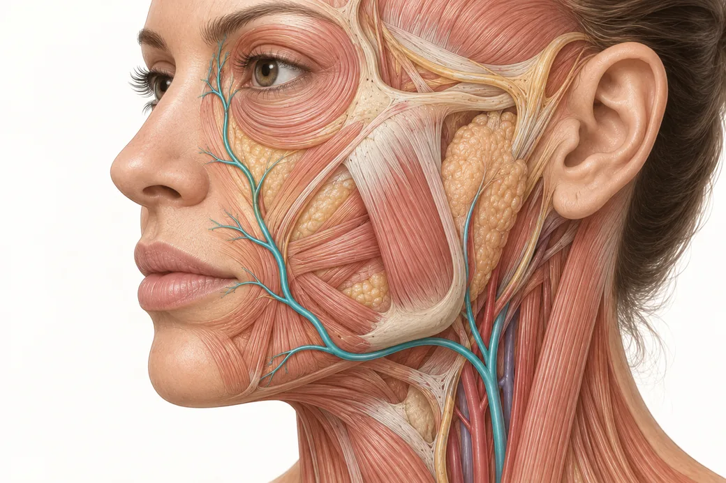 Clinical GuideAnastomoses: Why the Complication Doesn't Appear Where You InjectedFacial artery anastomoses explain why a lip treatment causes nasal skin changes. How arterial connections reframe injector triage and p
 Clinical GuideFiller Vascular Occlusion and Vision Loss: The Glabellar Crossroad, Step by StepHow filler vascular occlusion at the glabella causes vision loss — the retrograde embolic chain from the angular artery to the retina,
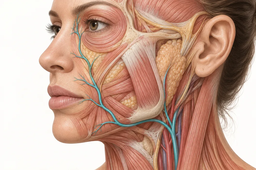 Clinical GuideFacial Artery Variability: Why Landmarks Are Probabilities, Not CertaintiesFacial artery variability in cadaver data — termination points, branch prevalence, tortuosity and depth, and what probabilistic anatomy
Train with Empire
This guide is clinical education. The technique behind it is taught hands-on, on live patients, with faculty beside you.
Explore Anatomical-Based Aesthetics Training →Disclaimer
This article reflects the clinical opinions and experience of Melissa Pulcini-Buttine, PA, an independent faculty member contributing to Empire Medical Training's curriculum. The views expressed are the author's own and do not necessarily represent those of Empire Medical Training.
It is professional education, not medical advice, and is no substitute for hands-on training or independent clinical judgment. Licensed clinicians remain responsible for their own patient selection, technique and outcomes, for verifying current product labelling, and for practising within their scope and applicable law. Empire Medical Training accepts no liability for reliance on this content.
 Claim Your Prize!