








Retrograde embolisation is the answer to a question that unsettles every injector who thinks about it properly: how does product placed in the nasal dorsum end up in the retina? The needle was nowhere near the eye. The distance is centimetres. Blood in that territory flows the wrong way for it.
Melissa Pulcini-Buttine, PA, compresses the mechanism into one sentence when she teaches it: "Injection pressure overcomes the local flow. Press slow." And then the consequence: "You could literally retrograde that product into another vessel that's communicating nearby and cause ischaemia."
This article is the long version of that sentence — the hemodynamics, with what has actually been measured. It is deliberately the why. The habits that follow from it are a separate spoke in this cluster, because an injector who understands the mechanism will derive most of those habits unprompted, and will apply them better than one who memorised a list.
Step one: you have to be inside the vessel
Nothing that follows happens unless the needle or cannula tip is intraluminal. This sounds obvious and it matters, because it locates the whole failure at the moment of placement rather than at the moment of pressure.
How easily does that happen? A fresh cadaver head perfusion model built to simulate physiologic blood pressure and flow through the carotid, ophthalmic and supratrochlear arteries attempted cannulation of the superficial branch of the supratrochlear artery. Cannulation succeeded in all six specimens. The same study measured that vessel as sitting a mean of 1.5 mm from the epidermal surface (Cho KH, Dalla Pozza E, Toth G, Bassiri Gharb B, Zins JE. Pathophysiology Study of Filler-Induced Blindness. Aesthet Surg J. 2019;39(1):96-106. PMID 29873688).
One and a half millimetres. That is within the range of a superficial dermal injection, and it is a vessel that connects to the ophthalmic artery.
Step two: your thumb is already beating the patient's blood pressure
Here is the finding that reorganises how injectors should think about pressure, and it comes from outside aesthetics entirely.
Investigators instrumented 71 intralesional corticosteroid injections performed by three fellowship-trained ophthalmologists and measured injection pressure in real time. In 63 of 71 injections the maximum pressure exceeded 100 mmHg, with a range of 18.65 to 842.18 mmHg, and the authors reported that "each surgeon produced injection pressures greater than the systemic arterial pressures of each patient." Their conclusion: "A sufficient volume of corticosteroid injected at high injection pressure would account for the embolization of corticosteroid particles into the ocular circulation from retrograde arterial flow" (Egbert JE, Paul S, Engel WK, Summers CG. Arch Ophthalmol. 2001;119(5):677-683. PMID 11346395).
Note the detail that makes this damning rather than merely interesting: this was a non-masked protocol. Experienced injectors who knew their pressures were being measured still could not keep them below arterial pressure.
Filler makes it worse, and for a reason rooted in the material rather than the operator. Hyaluronic acid gels are pseudoplastic — they have a yield stress that must be overcome before they flow at all. A bench study across four HA fillers of differing rheology and three needle diameters concluded that "irrespective of the injection force, the ejection pressure was likely to be higher than the vascular pressure at the time of entry into the vessel, rendering the injection dangerous" (Lee Y, Oh SM, Lee W, Yang EJ. J Cosmet Dermatol. 2021;20(5):1551-1556. PMID 33713373).
Follow that to its conclusion, because it is the single most useful thing in this article:
You cannot inject filler at a pressure below arterial pressure. The gel will not move. Every filler injection ever performed has been delivered at a pressure capable of driving product retrogradely up an artery. The only variable that has ever protected your patients is that the tip was not in one.
This reframes the whole subject. "Press slow" is not a technique for staying below the threshold — there is no achievable pressure below the threshold. Slow injection changes something else, and the something else is volume and time, which is where the rest of the mechanism lives.
Step three: the retrograde column
With the tip intraluminal and the pressure above systolic, product travels against flow. The question is how far.
The cadaver perfusion model above is the first human study to substantiate the mechanism directly. Product injected into the cannulated supratrochlear artery produced emboli demonstrated in the ophthalmic artery in three of six specimens, with a C-arm angiogram showing a cut-off sign in the ophthalmic artery. The authors concluded that "retrograde hyaluronic acid filler emboli to the ophthalmic artery could be produced by the cannulation of the supratrochlear artery." The average injection pressure reported as sufficient to inoculate the supratrochlear artery and retrogradely fill the ophthalmic artery back to the takeoff of the retinal artery was 166 mmHg — a pressure comfortably within the range that ordinary thumbs generate.
An honesty check that belongs in the same paragraph. A live-animal study injected methylene blue into the rabbit facial artery and compared living and dead animals. In dead rabbits, retrograde travel to the ophthalmic system occurred in 20 of 20. In living rabbits, it occurred in 1 of 20 (P < 0.05). The authors concluded that fillers can retrogradely enter the ophthalmic artery in vivo, "although the possibility is much lower in vivo than it is in corpses" (Zheng H, Qiu L, Liu Z, et al. Aesthetic Plast Surg. 2017;41(5):1222-1227. PMID 28432417).
That result should change how you read every cadaveric finding in this field, including the one above. Cadaver models systematically overstate how easily retrograde embolisation occurs, because there is no opposing pulsatile pressure. This is consistent with what clinicians actually observe: the event is rare, not routine, even though the pressure condition is met on every injection. Living arterial pressure is genuinely protective — just not reliably so.
Step four: how little volume it takes
The "keep boluses under 0.1 mL" rule has a real origin, and it is routinely misquoted. It does not come from a measurement of the ophthalmic artery.
Six fresh cadaver heads, twelve hemifaces, latex arterial injection: the measured volume of the supratrochlear artery from the glabella to the orbital apex was 0.04 to 0.12 mL, mean 0.085 mL, with a mean length of 51.75 mm and a mean radius of 0.72 mm. The authors concluded that "a bolus of this critical volume may lead to a significant adverse outcome" (Khan TT, Colon-Acevedo B, Mettu P, DeLorenzi C, Woodward JA. An Anatomical Analysis of the Supratrochlear Artery. Aesthet Surg J. 2017;37(2):203-208. PMID 27530765).
An independent study of 36 fresh cadaver hemifaces corroborated the order of magnitude, measuring the path from the supraorbital artery to the central retinal artery at 0.083 cm³ and from the supratrochlear at 0.089 cm³, and concluding that "injecting as little as 0.08 ml of HA into the facial branch is enough to cause central retinal artery embolism" (Zhang L, Pan L, Xu H, et al. Aesthetic Plast Surg. 2019;43(4):1054-1060. PMID 31006827).
The rule is now contested, and injectors should know that. A CT study of 80 ophthalmic arteries in 40 living patients calculated a mean ophthalmic artery volume of 0.16 mL, and stated: "The volume of the ophthalmic artery appears to be 0.2 mL rather than 0.1 mL as previously reported. In addition, it appears impractical to limit the volume of soft tissue filler bolus injections to 0.1 mL" (Li XR, Hong WJ, Luo SK, et al. Aesthet Surg J. 2023;43(9):1025-1032. PMID 36866393).
The accompanying commentary supplies the framing that survives both figures: "any volume greater than zero could potentially result in injection-related visual compromise" (Wu WTL. Aesthet Surg J. 2023;43(9):1033-1035).
So the honest position on volume is this. Small aliquots remain correct, and the reason is not that 0.1 mL is a safe threshold below which nothing happens. It is that the volume delivered before you notice something is wrong determines how much artery gets filled, and every increment of that volume extends the column further upstream.
Step five: the product does not stay in one piece
Injectors tend to picture the embolus as a plug — a discrete worm of gel occupying a length of vessel. In-vitro perfusion work shows something messier and clinically more important.
Hyaluronic acid gels fragment extensively on arterial inoculation, with particle sizes ranging from under 50 µm to over 1 mm, and the fragmentation pattern correlates most strongly with tan(δ), a rheological measure of how solid or fluid a gel behaves (Soares DJ, McCarthy AD. The Impact of Gel Parameters on the Dispersal and Fragmentation of Hyaluronic Acid Gel Fillers within an Artificial Model of Arterial Embolism. Gels. 2024;10(8):530. PMID 39195059).
Downstream, that matters enormously. A microvascular model using 200–1000 µm channels found that occlusion predominated in channels of 300 µm or smaller (P < .0001), and that gel behaviour tracked rheology: "solid gels fractured into ovoid embolic particles, whereas soft, high-tan δ gels formed filamentous, nonocclusive strands." The same study found 22G cannulas produced larger particles and higher occlusion rates than 27G (31% vs 17%, P = .025), and concluded that HA fillers "behave as deformable embolic particles that disperse distally… supporting a concurrent microembolic mechanism" (Soares DJ, Trudel S, Siperstein R, Kean TJ, McCarthy AD. Aesthet Surg J. 2025;46(1):65-75. PMID 40795834).
Two further properties compound it. HA gels swell by 100–700% by volume, so an embolus becomes more occlusive after placement rather than less. And the embolus changes composition: in a rat model, "the composition of the emboli caused by arterial hyaluronic acid-induced occlusion changed from pure hyaluronic acid to a hyaluronic acid-thrombus mixture" over time (Chen Y, Zhang YL, Luo SK. Plast Reconstr Surg. 2019;143(4):1088-1097. PMID 30921126).
That last finding is the mechanistic reason a delayed dissolution attempt is a different proposition from an immediate one. Hyaluronidase digests hyaluronic acid; it does not digest thrombus. What filler dissolve can achieve depends partly on how much of the obstruction is still made of the thing the enzyme works on.
Step six: the four dissemination patterns
The most useful model for "product where you didn't put it" describes four patterns of arterial dissemination (Soares DJ. Molecules. 2022;27(17):5398. PMID 36080164):
- Type I — the filler disperses fully and embolises distally; the main lumen clears. Favoured by low volume, low viscosity and a large vessel.
- Type II — no dispersal; a proximally occlusive plug. Favoured by higher viscosity and cohesivity and larger volume.
- Type III — distal dispersal plus a proximal plug expanding anterograde.
- Type IV — distal dispersal plus a proximal plug expanding retrograde, occluding upstream branches.
Types III and IV are described as likely the most common patterns, and the author states that "cerebroretinal injuries arise by default from type IV filler disseminations, in which an occlusive plug retrogradely extends toward the origin of the retinal artery and back into the internal carotid/cerebral arteries."
This model explains something the simple retrograde picture does not: why a single event can produce both distant catastrophic consequences and local skin ischaemia, and why the skin findings can appear in territory you never touched. Melissa describes exactly this presentation from the chairside: "You'll start to see changes in the skin, pain outside of the area that you injected in."
What the model gives the injector is a reason to expect trouble away from the needle. Once plunger pressure is released and antegrade flow returns, the territory at risk is not confined to the vessel you entered — which is why the assessment after any suspected event has to cover the whole distribution of that system, not the injection site.
What gauge buys you, and why it is time
One in-vitro study links all of this back to something you control at the moment of injection.
Using a bifurcated tubing model perfused at 70 mmHg with a dynamometer delivering constant force, investigators compared 27G and 30G needles across three fillers. With a 27G needle, product reached the bifurcation in 9 of 9 trials, with retrograde flow beginning at 3.33 seconds and the bifurcation reached at 8.44 seconds. With a 30G needle, only 3 of 9 reached the bifurcation, 4 of 9 showed no retrograde flow at all, retrograde flow began at 14 seconds, and the bifurcation was reached at 33.33 seconds (Scott G, Khonda M, Hsu T, et al. Plast Reconstr Surg Glob Open. 2023;11(9):e5270. PMID 37711726).
The authors' conclusion is counter-intuitive and worth sitting with: thinner needles are less likely to cause retrograde occlusion despite requiring more extrusion force, because the lower flow rate buys time to notice something is wrong and stop.
That is the mechanistic justification for the most under-rated safety instruction in aesthetics. Melissa puts it as "stop early, because it can escalate fast." What the model shows is that early is measured in seconds — the difference between a column reaching a bifurcation at 8 seconds and at 33 seconds is the entire window in which a human being can perceive resistance, perceive pain, perceive blanching, and lift their thumb.
You are not injecting below a dangerous pressure. You are managing how much product crosses how much artery before you notice.
What the mechanism implies
Three things follow directly from the hemodynamics, and they are stated here as consequences rather than as a protocol — the practical habits, including where the evidence for each is strong and where it is contested, are the subject of the companion spoke in this cluster.
Pressure control is not threshold management. Since every filler injection exceeds arterial pressure, slow delivery is not keeping you under a line. It is reducing flow rate, which reduces the volume delivered per unit of time and extends the time available to detect and abort.
Volume limits are about the length of artery filled, not a magic number. The 0.1 mL heuristic has a genuine cadaveric origin in the supratrochlear path and is now contested by living-subject imaging. Smaller aliquots remain right for a reason that survives the dispute.
Time is the resource. Every element of the mechanism — needle gauge, flow rate, aliquot size, willingness to stop — converts into seconds of margin before an embolic column reaches a branch point.
Melissa's question is the one to carry into the room: "What are those vessels connected to, and what can that flow if you're pressing too hard?" The hemodynamics say the pressure is always sufficient. The anatomy decides whether it matters.
The teaching framing in this article reflects Melissa Pulcini-Buttine's clinical practice as taught in Empire Medical Training's hands-on curriculum. Technique is learned under supervision; this article is educational and is not a substitute for training.
Injectors who want to build this on dissected anatomy will find it in Empire's anatomical based aesthetics training and special anatomical cadaver aesthetics training. Regional technique is covered in complete dermal filler training and the highest-risk territories in master eye and nose injection training. The separate question of toxin and visual risk is addressed in can Botox cause blindness.
Melissa Pulcini-Buttine, PA — physician assistant of two decades; professor of anatomy and physiology for ~14 years; faculty member, Empire Medical Training; founder of an aesthetics practice in Greenwich, Connecticut.
Related guides in this cluster
Part of Facial Vascular Anatomy for Injectors.
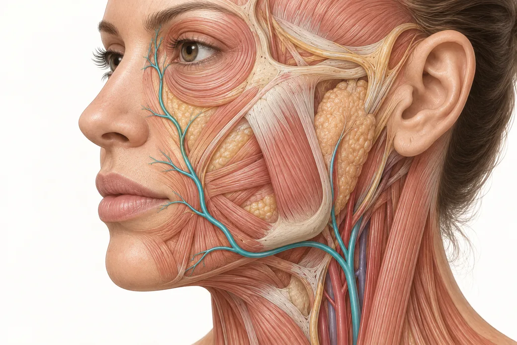 Clinical GuideAnastomoses: Why the Complication Doesn't Appear Where You InjectedFacial artery anastomoses explain why a lip treatment causes nasal skin changes. How arterial connections reframe injector triage and p
 Clinical GuideFiller Vascular Occlusion and Vision Loss: The Glabellar Crossroad, Step by StepHow filler vascular occlusion at the glabella causes vision loss — the retrograde embolic chain from the angular artery to the retina,
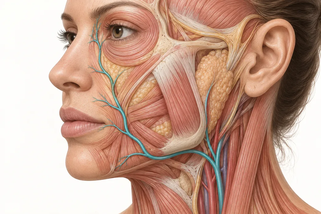 Clinical GuideFacial Artery Variability: Why Landmarks Are Probabilities, Not CertaintiesFacial artery variability in cadaver data — termination points, branch prevalence, tortuosity and depth, and what probabilistic anatomy
Train with Empire
This guide is clinical education. The technique behind it is taught hands-on, on live patients, with faculty beside you.
Explore Anatomical-Based Aesthetics Training →Disclaimer
This article reflects the clinical opinions and experience of Melissa Pulcini-Buttine, PA, an independent faculty member contributing to Empire Medical Training's curriculum. The views expressed are the author's own and do not necessarily represent those of Empire Medical Training.
It is professional education, not medical advice, and is no substitute for hands-on training or independent clinical judgment. Licensed clinicians remain responsible for their own patient selection, technique and outcomes, for verifying current product labelling, and for practising within their scope and applicable law. Empire Medical Training accepts no liability for reliance on this content.
 Claim Your Prize!