







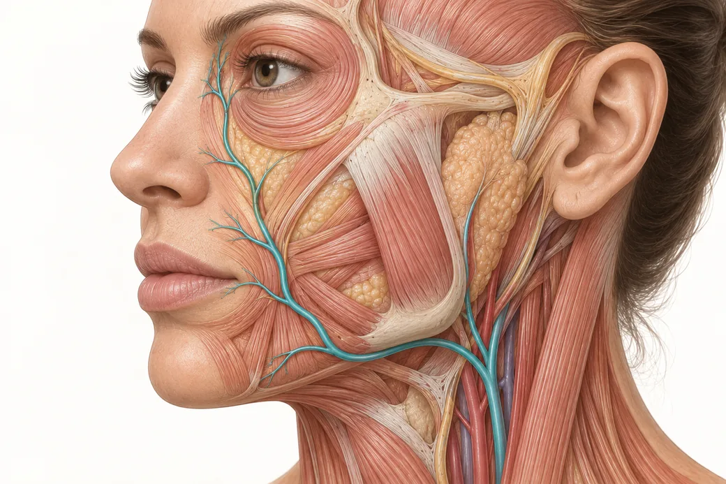
Knowing the name of a vessel is not where the story ends. You also have to know the facial layers — whether you are deep, middle or superficial, what occupies that plane in that region, and what is immediately above and below you.
A vascular map with only two dimensions is a map of a photograph. The face is a solid. Every artery on the map runs at a depth, in a layer, and the layer it occupies changes as it travels. Add that third dimension and the map becomes usable; leave it out and you have a set of coordinates with no elevation data.
This page is descriptive anatomy. It covers what the layers are, which structures live in each, and how the arteries relevant to injectors distribute across them. It does not prescribe planes for particular products in particular regions — that is a technique decision made under supervision, and it is not what this reference is for.
The five layers
The face is organised into five concentric layers, consistent in sequence from the scalp to the neck even though their individual character changes markedly by region (Cotofana et al., JDDG, 2019).
Layer 1 — Skin. Epidermis and dermis. Thickness varies substantially by region: thin and mobile over the eyelid, thick and sebaceous over the nasal tip.
Layer 2 — Subcutaneous fat. The superficial fat compartments, held in a scaffold of fibrous septae — the retinacula cutis — that run between the dermis above and the fascia below. This layer is compartmentalised rather than continuous, and the compartments have boundaries.
Layer 3 — The superficial musculoaponeurotic system (SMAS). A continuous fibromuscular sheet that is aponeurotic in some regions and frankly muscular in others — it is continuous with the galea above, the platysma below, and it invests the mimetic muscles. In much of the midface, "layer 3" and "the muscle" are the same thing.
Layer 4 — Deep fat and the areolar plane. The deep fat compartments and the loose areolar spaces that allow the superficial layers to glide over the deep ones. This layer is where the face's mobility comes from.
Layer 5 — Periosteum and deep fascia. The final layer over bone, and the deep fascia over structures such as the parotid and the masseter.
Two structural points matter more than the list itself.
The facial fat lives in layers 2 and 4 — never in layer 3. When you read about superficial versus deep fat compartments, you are reading about two different layers separated by a fibromuscular sheet, not about two depths of the same tissue.
The retaining ligaments run perpendicular through all of it. True retaining ligaments — orbital, zygomatic, buccal-maxillary and mandibular — originate from periosteum or deep fascia and travel straight up through the layers to insert into the dermis. They anchor the soft tissue to the skeleton at defined locations. Neurovascular structures often travel with, or close to, these vertical routes.
Why layers, and not millimetres
Here is the argument for the whole framework in one comparison.
Three millimetres at the mandibular border, where the facial artery enters the face at roughly 6 mm depth, is safely superficial to the trunk. Three millimetres at the medial canthus, where the terminal angular artery sits around 1 mm from the skin, is below the vessel. Same number on the ruler. Opposite anatomical situations.
A millimetre reading tells you how far your needle went. A layer tells you what your needle is in. Only the second one is clinically interpretable, and the conversion between them changes from region to region.
That is why experienced anatomy teaching insists on plane language — subdermal, subcutaneous, intramuscular, submucosal, supraperiosteal — rather than numbers. The number is an output; the plane is the decision.
The layers are not the same thickness everywhere
The model is consistent in sequence, not in dimensions, and the regional differences are exactly where injectors get caught.
Over the glabella and central forehead, the total soft tissue envelope is thin. Layer 2 is sparse, layer 3 is frontalis and corrugator, and the periosteum is close. There is very little vertical room between planes.
Over the mid-cheek, the envelope is at its most generous — well-developed superficial and deep fat compartments, a distinct SMAS, and a wide areolar plane. The layers are furthest apart here, which is why the cheek tolerates plane error better than the glabella does.
Over the lips, the sequence is modified entirely. There is no SMAS in the vermilion. The orbicularis oris is the muscular layer, there is a submucosal plane internally, and the soft tissue depth from vermilion to mucosa is a matter of a few millimetres in total.
Over the nasal dorsum and tip, the skin is thick and adherent, the subcutaneous layer is thin, and the vessels run in a plane that is superficial to the nasal skeleton but deep to the dermis — a narrow corridor with very little margin on either side.
So "superficial" is not a fixed depth. It is a position within a stack whose height changes across the face.
Where the arteries actually sit
This is where the layer model earns its keep, because published measurements let us place specific vessels in specific planes.
The facial artery trunk runs deep where it enters the face and becomes more superficial as it ascends — roughly 6 mm at the mandibular origin, 8–9 mm at the level of the oral commissure, 9–10 mm at the nasal ala on pooled data (Trzeciak et al., Aesthetic Plastic Surgery, 2025). In the region of the nasolabial fold it commonly runs in the subcutaneous plane — layer 2 — rather than deep, which is the reverse of what its mandibular depth would lead you to expect.
The labial arteries were measured by ultrasound in 41 living volunteers: mean depth 5.6 ± 0.13 mm for the superior labial artery in the upper lip and 5.2 ± 0.14 mm for the inferior labial in the lower lip. More usefully, they were located in the submucosal plane in 58.5% of measurements, intramuscular in 36.2%, and subcutaneous in only 5.3% (Cotofana et al., Aesthetic Surgery Journal, 2020;40(12):1327–1335). Both arteries were found within the red lip rather than above the vermilion border in the large majority of cases — 83% of upper lips and 86.2% of lower lips.
That is a layer distribution, not a depth. It says the labial arteries are predominantly deep to the muscle relative to the skin surface, sitting toward the wet side of the lip, with only a small minority running in the subcutaneous plane.
The supratrochlear artery in the glabella and central forehead runs subcutaneously through most of its course: its superior third beneath the dermis and above the fat, its inferior two-thirds above the frontalis and beneath the fat, with a terminal muscular branch passing under the frontalis. Central forehead ultrasound measured its superficial branch at 4.2 mm and a deep branch at 5.9 mm (Phumyoo et al., Clinical Anatomy, 2020). A vessel that spends most of its course in layer 2 of a region where layer 2 is thin is a vessel with very little cover.
The angular artery at the medial canthus arrives at roughly 1 mm from the skin surface, and in vivo work on the angular segment reports it running superficial to the levator labii superioris alaeque nasi in the large majority of cases. Superficial to the muscle — meaning that in this territory, the muscle is not between the vessel and your needle.
Notice what these four descriptions have in common: no vessel stays in one layer along its course. The facial artery is deep at the mandible, subcutaneous at the fold. The supratrochlear artery is subcutaneous in the forehead and intramuscular at its terminal branch. Layer membership is a function of position, exactly as depth is.
What the layers tell you about spread
One more descriptive property, because it explains observations injectors make constantly without a framework for them.
Layers 2 and 4 are compartmentalised — the fat sits in bounded compartments with septal walls. Layer 4's areolar spaces are glide planes, which means material placed there encounters comparatively little resistance in the plane and considerable resistance crossing out of it. Layer 3 is a continuous sheet, and structures that penetrate it — perforating vessels, the retaining ligaments — do so at defined points rather than anywhere.
The practical consequence is descriptive rather than prescriptive: material introduced into a bounded compartment behaves differently from material introduced into a glide plane, and both behave differently from material introduced into a septal or ligamentous route. Where a product ends up is determined by the layer's architecture as much as by where the needle tip was. That is also why volume placed in one compartment can produce a contour change with boundaries that surprise you — the boundaries belong to the anatomy, not to the injection. For the broader picture of how these compartments change with age, see facial volume loss.
What this page deliberately does not do
It does not tell you which plane to use for which product in which region.
That is a genuine boundary, not modesty. Plane selection is a decision made with a specific product's rheology, a specific patient's anatomy, a specific goal and a specific delivery tool, and it is learned under supervision with your hands in tissue. Reading a table of recommended planes produces injectors who can recite a plane and cannot recognise one.
What this reference gives you is the vocabulary and the anatomy to make that decision meaningfully, and to understand what a supervisor means when they say "you're too superficial here." The layer model is a way of seeing. The plane you choose is a clinical judgement built on top of it.
Learning layers in tissue
Layers are the part of facial anatomy that is genuinely hard to learn from images, because the defining property of a layer is what it feels like to pass through it and what separates it from the one above. A diagram renders five bands of colour. Tissue renders a fibrous septum, a glide plane that opens with almost no resistance, an aponeurosis that resists, and a periosteum that stops you.
That difference is why Empire Medical Training's Special Anatomical Cadaver Aesthetics Training and Anatomical Based Aesthetics Training work layered dissection directly rather than treating depth as a number to be memorised.
These anatomical relationships reflect Melissa Pulcini-Buttine's teaching in Empire Medical Training's hands-on curriculum, supported by the published literature cited above. This article is educational and is not a substitute for supervised training.
Melissa Pulcini-Buttine, PA — physician assistant of two decades; professor of anatomy and physiology for ~14 years; faculty member, Empire Medical Training; founder of an aesthetics practice in Greenwich, Connecticut.
Related guides in this cluster
Part of Facial Vascular Anatomy for Injectors.
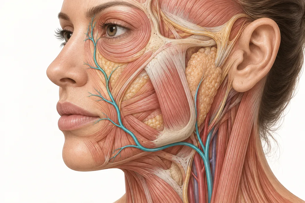 Clinical GuideAnastomoses: Why the Complication Doesn't Appear Where You InjectedFacial artery anastomoses explain why a lip treatment causes nasal skin changes. How arterial connections reframe injector triage and p
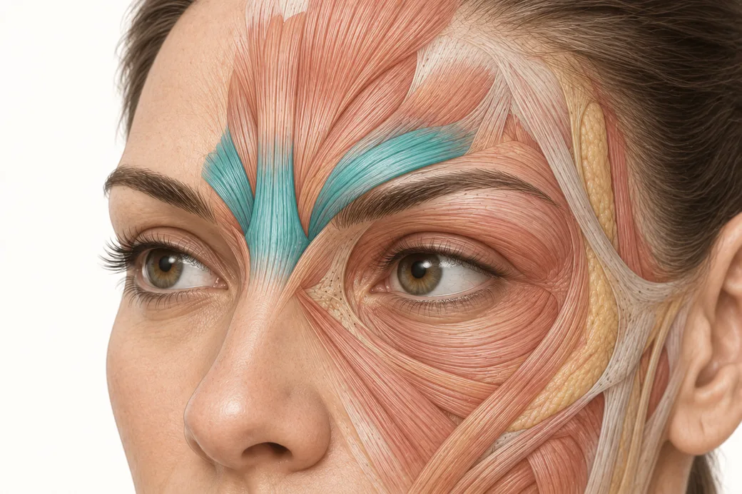 Clinical GuideFiller Vascular Occlusion and Vision Loss: The Glabellar Crossroad, Step by StepHow filler vascular occlusion at the glabella causes vision loss — the retrograde embolic chain from the angular artery to the retina,
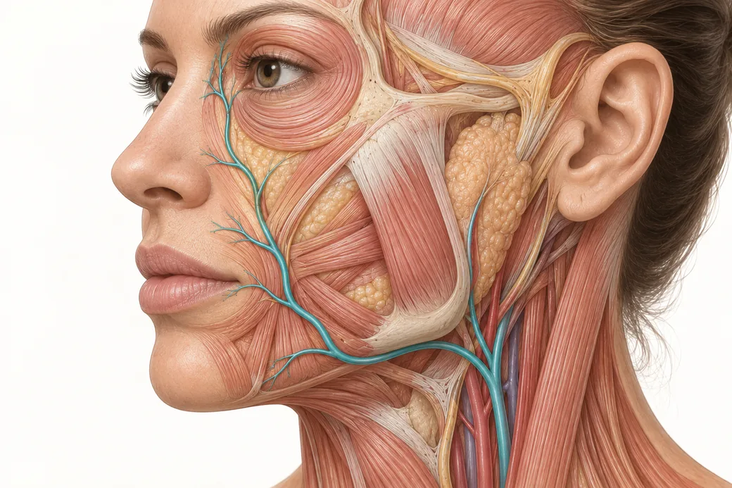 Clinical GuideFacial Artery Variability: Why Landmarks Are Probabilities, Not CertaintiesFacial artery variability in cadaver data — termination points, branch prevalence, tortuosity and depth, and what probabilistic anatomy
Train with Empire
This guide is clinical education. The technique behind it is taught hands-on, on live patients, with faculty beside you.
Explore Anatomical-Based Aesthetics Training →Disclaimer
This article reflects the clinical opinions and experience of Melissa Pulcini-Buttine, PA, an independent faculty member contributing to Empire Medical Training's curriculum. The views expressed are the author's own and do not necessarily represent those of Empire Medical Training.
It is professional education, not medical advice, and is no substitute for hands-on training or independent clinical judgment. Licensed clinicians remain responsible for their own patient selection, technique and outcomes, for verifying current product labelling, and for practising within their scope and applicable law. Empire Medical Training accepts no liability for reliance on this content.
 Claim Your Prize!