







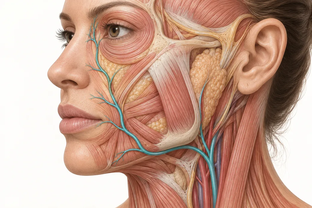
Facial artery variability is the reason a vascular map is useful and the reason it can never be trusted the way a road map is trusted. The facial artery has more anatomical variation than almost any vessel an injector works around. It can be markedly tortuous. It changes depth along its course. It can end early, run duplicated, or be so hypoplastic that another vessel has taken over its territory.
Most injectors have heard that. Far fewer have seen the numbers, and the numbers are what convert a vague caution into a working model. What follows is what the dissection, CT-angiography and ultrasound record actually reports — and what a clinician should do with anatomy that comes with a confidence interval.
What the dissection record shows
The most useful recent dataset comes from a cadaveric study of 52 cadavers yielding 102 hemifaces (Nguyen et al., Archives of Craniofacial Surgery, 2024;25(2):77–84). In those 102 facial arteries, the investigators documented eight distinct termination points and 35 different branching-pattern combinations.
Thirty-five. Not thirty-five minor deviations from a norm — thirty-five combinations of which branches were present and in what order they arose.
That single figure should change how you read any facial artery diagram, including the one in this cluster's pillar. A diagram shows one arrangement. The population contains dozens.
Termination: the vessel frequently does not end where you were taught
The textbook account is that the facial artery ascends and terminates as the angular artery at the medial canthus. In the 102-hemiface series, the termination points broke down like this:
| Termination point | Prevalence |
|---|---|
| Lateral nasal artery | 32.35% |
| Angular artery | 30.39% |
| Duplex type | 17.65% |
| Inferior alar artery | 6.86% |
| Superior labial artery | 4.90% |
| Inferior labial artery | 4.90% |
| Forehead branch | 1.96% |
| Hypoplastic (short course) | 1.0% |
Read the bottom half of that table carefully. In roughly one hemiface in ten, the facial artery ends at the lip — it terminates as the superior or inferior labial artery and never reaches the nose at all. In another 7%, it ends at the nasal base. In a small fraction it is essentially absent as a functional midface vessel.
A meta-analysis pooling the wider literature reports that termination as either the lateral nasal or the angular artery accounts for about 69.81% of facial arteries (Koziej et al., Clinical Anatomy, 2022). Which means that in close to a third of cases, the vessel ends somewhere else.
The clinical reading is not "the map is wrong." It is: the territory a given artery supplies in this patient may be smaller, or differently shaped, than the diagram suggests — and something else is supplying the remainder. When a facial artery terminates early, the midface and nose are perfused by collateral inflow, often from the ophthalmic system descending from above. That patient's midface is not less vascular. It is differently vascular, and possibly more directly connected to the orbit.
Branch presence: what you may and may not assume
From the same 102 hemifaces:
- Superior labial artery: 87.25% — the most consistently present of the named branches, and still absent in roughly one in eight
- Inferior labial artery: 78.43% — absent in roughly one in five
- Angular artery: 33.33% — present in only one hemiface in three
A separate classification of the superior labial artery divides it into four types: the facial artery independently giving rise to both the superior labial and an alar branch (56.7%), the alar branch arising from the superior labial (21.7%), the facial artery terminating as the superior labial (15.0%), and the superior labial absent altogether (6.7%).
The absence figures are the interesting ones, and they are frequently misread. A missing superior labial artery does not mean an avascular lip. It means the lip is supplied by a different arrangement — a contralateral vessel crossing the midline, an enlarged counterpart, or a branch from a neighbouring territory. Variability redistributes vasculature; it does not remove it. An injector who reasons "the textbook vessel probably isn't here, so this is safer" has drawn exactly the wrong conclusion.
Tortuosity, and the nasolabial fold problem
Tortuosity is the variable that makes surface landmarks least reliable, because a tortuous vessel can cross a landmark repeatedly or run parallel to it a few millimetres away, and the surface gives you no way to tell which.
The nasolabial fold is the worked example. Yang and colleagues found the facial artery crossed the nasolabial fold in 33.9% of cases and ascended within 5 mm of it in 42.9% (Plastic and Reconstructive Surgery, 2014;133(5):1077–1082). So in roughly three-quarters of faces, the vessel is either on the fold or within five millimetres of it — and in the remaining quarter it is somewhere else entirely.
A cadaveric CT-angiography study of 52 cadavers put the horizontal relationship between the artery and the fold at between roughly −1.90 ± 2.40 mm and −6.92 ± 3.70 mm depending on the level measured. Look at those standard deviations rather than the means. A mean offset of 1.9 mm with a standard deviation of 2.4 mm describes a vessel whose position, in a given individual, could plausibly be on either side of the landmark.
That is what "the landmark is a probability" means quantitatively. The centre of the distribution is useful. The width of the distribution is the part that hurts you.
Depth is not one number
The second axis of variation is depth, and it varies both along the vessel and between individuals.
A 2025 meta-analysis of twelve studies measured facial artery depth at five defined levels and reported (95% CI): 5.98–6.62 mm at the mandibular origin, 8.36–9.20 mm at the level of the oral commissure, and 9.52–10.51 mm at the level of the nasal ala (Trzeciak et al., Aesthetic Plastic Surgery, 2025; doi:10.1007/s00266-025-04833-9). The same analysis noted that the depth between the nasal ala and the oral commissure varies significantly — i.e. the segment in the middle of the midface is the least predictable one.
Contrast that with the terminal angular segment, which ultrasound places at approximately 1 mm from the skin surface at the medial canthus. The same named vessel runs at roughly six millimetres where it enters the face and about one millimetre where it ends.
Two consequences follow, and they point in opposite directions, which is why depth cannot be treated as a simple rule.
Deeper is not automatically safer. Around the nasolabial fold, the facial artery commonly runs in the subcutaneous plane, and published discussion of that region suggests supraperiosteal placement may be safer than injecting into the SMAS layer there. At the level of the oral commissure, with the trunk sitting 8–9 mm down, injection to a depth of about 4.5 mm — superficial to the artery — has been described as comparatively safe. The safe plane is region-specific, and in some regions the safer plane is the deep one while in others it is the superficial one.
A depth measurement is not a location. Three millimetres at the mandibular border, three millimetres at the commissure and three millimetres at the medial canthus put your needle tip in three entirely different anatomical situations. The layer concept — which is the subject of its own spoke in this cluster — is what makes depth interpretable.
Why the published figures disagree, and why that is informative
Here is a discrepancy worth sitting with rather than smoothing over.
The 102-hemiface dissection series reports an angular artery present in 33.33% of specimens. A cadaveric CT-angiography study reports the facial artery continuing as an angular artery in 83.7% of specimens. Those cannot both be describing the same thing.
They are not. The difference is largely definitional: some authors reserve the name "angular artery" for a vessel that reaches the medial canthus, while others apply it to any continuation of the facial artery past the level of the nasal ala. Methodology contributes too — gross dissection, latex or lead-oxide perfusion with CT reconstruction, and Doppler ultrasound in living subjects each detect small vessels differently, and living tissue under normal perfusion pressure does not look like fixed cadaveric tissue. Study populations differ in ancestry and age. Prevalence ranges across the literature reflect all of this: roughly 32% to 74% for the angular artery, 77.5% to 98.0% for the superior labial artery, and 57.5% to 100% for the inferior labial artery.
The temptation is to pick the number you like and cite it. Resist it. The honest reading is that the range is the finding. Where studies converge — the superior labial artery is usually present, the facial artery usually gets to the nose or the canthus, the vessel gets shallower as it ascends — you can lean on the consensus. Where they diverge by a factor of two, that divergence is telling you the structure is genuinely inconstant, and the correct clinical response to an inconstant structure is margin.
Asymmetry within the same patient
One more variable that gets lost in population statistics: the two sides of a single face are not required to match.
Differences in branching pattern, termination point, depth and course between left and right hemifaces of the same cadaver are routinely documented — which is why studies count hemifaces rather than cadavers in the first place. The 102-hemiface series is 52 cadavers precisely because each side is treated as an independent observation.
Practically: confirming a landmark on one side tells you very little about the other. If you palpate the facial artery pulse at the antegonial notch on the right, you have learned about the right side.
How to act on a probability
If landmarks are probabilistic, what is left? Quite a lot, but it is a different kind of knowledge than most injectors expect.
Use the landmark as a prior, not a fact. The antegonial notch tells you where the facial artery enters the face with high reliability, because that crossing is anatomically constrained by bone. Almost nothing downstream of it is that reliable. Weight your confidence accordingly.
Palpate what can be palpated. The facial artery pulse at the mandibular border is real, patient-specific information, available in seconds, on the side you are about to treat.
Build margin where the distribution is widest. The segment between the oral commissure and the nasal ala is where the depth data are least consistent and the tortuosity greatest. That is where conservative volume, slow delivery and deliberate plane selection buy the most.
Let variability drive technique choice, not just site choice. Product, plane, needle versus cannula, injection speed and volume are all adjustable in response to how uncertain the anatomy is in that region.
Be honest about what ultrasound does and does not solve. Doppler ultrasound genuinely images the vessel in the patient in front of you, and it is the only tool that converts a population probability into an individual observation. It is also operator-dependent, adds time, is not universally available, and images the vessel at the moment of scanning rather than continuously during a procedure. It narrows uncertainty considerably. It does not eliminate it, and published ultrasound work on the midface angular artery concludes explicitly that there is no guaranteed safe location in that region (Cotofana et al., Aesthetic Surgery Journal, 2021;41(7):805–813).
Why this is an argument for dissection
You can read this page and retain the numbers for a week. What changes an injector permanently is opening ten faces and finding ten different arteries — a vessel that stops at the lip, a vessel that crosses the fold twice, a left side that does not match the right.
That is the specific value of cadaveric work, and it is why Empire Medical Training teaches Special Anatomical Cadaver Aesthetics Training alongside Anatomical Based Aesthetics Training, and why the anatomy precedes the technique in Complete Dermal Filler Training. In a cadaver lab you do not learn that the facial artery is variable. You experience it, and you stop expecting the diagram.
These anatomical observations reflect Melissa Pulcini-Buttine's teaching in Empire Medical Training's hands-on curriculum, supported by the published literature cited above. This article is educational and is not a substitute for supervised training.
Melissa Pulcini-Buttine, PA — physician assistant of two decades; professor of anatomy and physiology for ~14 years; faculty member, Empire Medical Training; founder of an aesthetics practice in Greenwich, Connecticut.
Related guides in this cluster
Part of Facial Vascular Anatomy for Injectors.
 Clinical GuideAnastomoses: Why the Complication Doesn't Appear Where You InjectedFacial artery anastomoses explain why a lip treatment causes nasal skin changes. How arterial connections reframe injector triage and p
 Clinical GuideFiller Vascular Occlusion and Vision Loss: The Glabellar Crossroad, Step by StepHow filler vascular occlusion at the glabella causes vision loss — the retrograde embolic chain from the angular artery to the retina,
 Clinical GuideThinking in Layers: Depth as the Second Axis of the Vascular MapFacial layers explained for injectors — the five-layer model, which layer each facial artery occupies, and why a depth in millimetres m
Train with Empire
This guide is clinical education. The technique behind it is taught hands-on, on live patients, with faculty beside you.
Explore Anatomical-Based Aesthetics Training →Disclaimer
This article reflects the clinical opinions and experience of Melissa Pulcini-Buttine, PA, an independent faculty member contributing to Empire Medical Training's curriculum. The views expressed are the author's own and do not necessarily represent those of Empire Medical Training.
It is professional education, not medical advice, and is no substitute for hands-on training or independent clinical judgment. Licensed clinicians remain responsible for their own patient selection, technique and outcomes, for verifying current product labelling, and for practising within their scope and applicable law. Empire Medical Training accepts no liability for reliance on this content.
 Claim Your Prize!