








Melissa Pulcini-Buttine, PA, ends her teaching on danger zones with four instructions delivered as a single unit: "Aspirate, pressure low, smaller volumes, stop early — because it can escalate fast."
They are the right four habits to reduce vascular complications, and each rests on a different body of evidence. Two carry strong guideline backing and solid mechanistic data. One is supported by mechanism and consensus. And one — aspiration — sits inside an active dispute in the peer-reviewed literature, with major consensus documents on opposite sides.
This article sets out what stands behind each of them. Where the evidence is strong it says so. Where the field is genuinely divided — as it is on aspiration — it lays out both cases rather than picking the one that makes the cleaner article.
Habit 1: Low pressure and slow delivery — strongest evidence
This is the habit with the clearest guideline support.
The Soft-Tissue Filler Safety recommendations grade it as a strong recommendation with moderate certainty of evidence: "Inject slowly with low plunger pressure, using small volumes with each pass, while keeping the cannula or needle moving" (Jones DH, Fitzgerald R, Cox SE, et al. Preventing and Treating Adverse Events of Injectable Fillers: Evidence-Based Recommendations From the American Society for Dermatologic Surgery Multidisciplinary Task Force. Dermatol Surg. 2021;47(2):214-226. PMID 33543879).
Melissa's version — "injection pressure overcomes the local flow, press slow" — states the mechanism correctly. The detailed hemodynamics are set out in the companion spoke on retrograde embolisation; the short version is that filler ejection pressure reliably exceeds arterial pressure, so slow delivery is not keeping you below a threshold. It is reducing flow rate, which buys detection time.
How much time is measurable. In a perfused bench model at constant force, a 27G needle produced retrograde flow at 3.33 seconds and reached a vessel bifurcation in all nine trials at 8.44 seconds; a 30G needle produced no retrograde flow at all in four of nine trials and took 33.33 seconds to reach the bifurcation (Scott G, Khonda M, Hsu T, et al. Plast Reconstr Surg Glob Open. 2023;11(9):e5270. PMID 37711726). Slower flow is more seconds in which a human can notice and abort.
One genuine unresolved conflict belongs here, because it is the kind of thing that gets papered over. Cadaveric work found an average injection pressure of 166.7 mmHg (range 160-180) sufficient to drive filler retrogradely from the supratrochlear artery into the ophthalmic artery (Cho KH, Dalla Pozza E, Toth G, Bassiri Gharb B, Zins JE. Aesthet Surg J. 2019;39(1):96-106. PMID 29873688), and a rheology study concluded that ejection pressure likely exceeds vascular pressure regardless of injection force (Lee Y, Oh SM, Lee W, Yang EJ. J Cosmet Dermatol. 2021;20(5):1551-1556. PMID 33713373).
Against that, a force study reached the opposite conclusion: "typical injection pressures generated by experienced injectors were significantly lower than that required to cause propagation of filler… and significantly lower than mean arterial pressure" (Ramesh S, Le A, Katsev B, Ugradar S. The Force Required to Inject a Column of Filler Through Facial Arteries. Dermatol Surg. 2020;46(9):e32-e37. PMID 31688236).
These cannot both be right, and the disagreement has not been settled. It does not change the recommendation — slow and low-pressure is graded strongly on other grounds — but injectors should know that the confident mechanistic story they have been told rests on findings that conflict.
Habit 2: Small volumes and small aliquots — strong evidence
Also strongly supported, and with a striking recent bench result.
Consensus guidance on visual loss states it as a numbered recommendation: "Micro-boluses should be injected in small aliquots (<0.1 mL)" (Goodman GJ, Magnusson MR, Callan P, et al. Aesthet Surg J. 2020;40(9):1009-1021. PMID 31693068). The Task Force recommendation quoted above carries the same instruction inside its strong grading.
The mechanism is not subtle. In a perfusion model comparing a 1 mL bolus against a 0.2 mL bolus, occlusion occurred in 57.1% versus 16.0% (P < 0.0001) and retrograde flow in 100% versus 10% (P = 0.005) (Soares DJ, Wu J, McCarthy AD. Aesthet Surg J. 2026, online ahead of print. PMID 42481041).
The reason the threshold sits where it does is anatomical. Cadaveric measurements of the arterial path from the glabellar region toward the orbit cluster between roughly 0.04 and 0.09 mL, depending on the study and the vessel measured — meaning a bolus in that range is capable of filling the relevant segment. That figure and its recent challenge from living-subject imaging are examined in the retrograde embolisation spoke; the practical point for this article is that small aliquots are right for a mechanistic reason that survives the dispute about the exact number.
A corollary that injectors under-apply: the Task Force wording is "small volumes with each pass, while keeping the cannula or needle moving." A moving tip cannot deposit a column into a fixed intraluminal position. This matters later, because it sits in direct tension with the fourth habit.
Habit 3: Stop early — mechanism and consensus
Melissa's framing is "stop early, because it can escalate fast," and the mechanism supports her: the Scott data above show that the difference between an embolic column reaching a branch point at 8 seconds and at 33 seconds is the entire window in which a person can react.
Two practical points determine whether an injector can actually use that window.
The signal to stop is often not pain. In a multinational survey of experienced injectors reporting intravascular events, the initial signs were minor livedo in 63%, pallor in 41% and pain in 37% — and pain was mild or absent in 47% of events (Goodman GJ, Roberts S, Callan P. Aesthetic Plast Surg. 2016;40(4):549-555. PMID 27286849). An injector waiting for the patient to flinch will miss roughly half of these. The classic findings to watch for are "skin blanching, livedo reticularis, slow capillary refill, and dusky blue-red discoloration" (DeLorenzi C. Aesthet Surg J. 2014;34(4):584-600. PMID 24692598).
The decision has to be pre-committed. In the moment, every early sign is ambiguous — blanching can be pressure blanch, resistance can be a fibrous septum, pain can be a sensitive patient. An injector who has not decided in advance what will make them stop will resolve each ambiguity in favour of continuing.
The support for this habit is mechanistic and consensus-based rather than outcome-based: the bench data establish the seconds available, and the guidelines instruct the injector to abort on the early signs. That is the right basis on which to act, and it is exactly why the decision has to be pre-committed rather than made in the moment.
Habit 4: Aspiration — genuinely contested
Melissa teaches aspiration emphatically and twice. "Besides knowing your anatomy you also need to aspirate. That's another safety mechanism. If you have a positive aspiration, do not push any product, because then you can spread it to the vessels that anastomose to something dangerous." And in her closing: "Always aspirate before you press anything."
She is in substantial and credentialed company. Aspiration is also one of the few manoeuvres in injectable practice on which the major consensus documents themselves diverge, and an injector should understand what sits behind each position rather than be handed either camp's as settled fact.
The case for aspiration
The foundational paper tested withdrawal through syringes containing 0.1 mL of each of 17 filler products. Aspiration was negative with 8 products (47%) and positive with 9 (53%) — and critically, every product that tested negative converted to positive when a larger-gauge needle was used. In a rabbit ear vein, all five products tested aspirated positive (Casabona G. Blood Aspiration Test for Cosmetic Fillers to Prevent Accidental Intravascular Injection in the Face. Dermatol Surg. 2015;41(7):841-847. PMID 26079591).
Positive aspirations happen in real practice, and a descriptive analysis of 213 of them estimated an incidence of 0.04% to 0.9% of injections, found that aspiration was achievable in under two seconds with an appropriate product and needle pairing, and found that primed needles produced significantly more positives than unprimed (P < 0.001) (Tseng FW, Bommareddy K, Frank K, et al. Aesthet Surg J. 2021;41(5):616-624. PMID 32504468).
The largest real-world dataset — 5,106 aspirations across 1,007 patients in 14 practices in nine countries, using a slow pull-back held at least five seconds — found 35 positives, a rate of 0.69%, with a between-investigator range of 0% to 6.72% (Nikolis A, Metelitsa A, Avelar L, et al. Aesthet Surg J. 2025;45(12):1285-1290. PMID 40729512).
The technical objections are partly answerable. Priming the needle with saline or air rather than filler dramatically improves detection: one study found plain aspiration succeeded "only in a few trials" at 27G and failed in all 29G trials, while saline-flashing the needle first gave 100% success in one to two seconds across all gauges and six fillers (Kogan I, Korolik P, Cartier H, Adhoute H, Liberzon A. J Cosmet Dermatol. 2020;19(10):2513-2518. PMID 32749775). A separate prospective model found saline- or air-primed needles flashed immediately in all 48 such tests (P < 0.0001).
An expert consensus explicitly endorses the practice, recommending that "aspiration should be performed as a prophylactic measure, particularly in highly vascularized areas, and a new needle without filler should be used prior to deep bolus injections… at least 0.1-0.2 cc of air, and waiting for at least 4 s" (Urdiales-Gálvez F, Delgado NE, Figueiredo V, et al. Aesthetic Plast Surg. 2017;41(3):667-677. PMID 28411354).
The case against
The opposing consensus is equally blunt. Recommendation 9 of the visual-loss consensus reads, verbatim: "There is currently no evidence to support aspiration as a safety measure" (Goodman et al. 2020, cited above).
The sensitivity data are the core of the argument. Testing 24 fillers across 11 needle sizes against a blood bag pressurised to 150 mmHg — 340 tests — found only 33% positive at one second and 63% at ten seconds overall (37% and 74% respectively with manufacturer-supplied needles) (Van Loghem JA, Fouché JJ, Thuis J. Sensitivity of aspiration as a safety test before injection of soft tissue fillers. J Cosmet Dermatol. 2018;17(1):39-46. PMID 28987016). Even at a ten-second pull, between a quarter and a third of intravascular placements produced no flashback.
An in-vitro evaluation found that no filler exhibited flash below a two-second pull, some required over ten seconds, some required twenty, and one did not flash at all. In fairness to that paper — and this is the kind of thing that gets lost when a study is recruited to an argument — its own stated conclusion is that pre-injection aspiration "may have utility as a safety checkpoint." It is not an anti-aspiration paper (Torbeck RL, Schwarcz R, Hazan E, et al. Dermatol Surg. 2019;45(7):954-958. PMID 30624257).
A physiologically pressurised model with animal correlation produced the single most alarming combination: a 30G needle primed with a high-G′ volumising filler was negative on all in-vitro arterial trials, all venous trials, and all in-vivo rabbit trials within a twenty-second cutoff. The same filler through a 27G needle flashed in about one second. The authors also note a mundane practical constraint: on most 1 mL filler syringes, the maximum achievable pullback is around 0.2 mL, not the larger volumes some protocols assume (Moon HJ, Lee W, Kim JS, Yang EJ, Sundaram H. Aesthet Surg J. 2021;41(8):NP1073-NP1083. PMID 33861847).
The harm argument, which is the part most injectors have not heard
The strongest objection is not that aspiration fails to detect. It is that the manoeuvre may invalidate its own result.
Under real-time ultrasound, aspiration produced retrograde needle-tip movement in every case, ranging from 1.1 to 5.3 mm with a mean of 2.9 mm — a displacement the authors describe as "of a magnitude consistently significant relative to the typical size of facial vessels." Double-handed technique performed better than single-handed (P = .037), and greater experience reduced movement (P < .0001) (Lin F, Goodman GJ, Magnusson M, et al. Movement of the Syringe During Filler Aspiration: An Ultrasound Study. Aesthet Surg J. 2022;42(10):1109-1116. PMID 35348575).
If the tip moves by an average of 2.9 mm during the pull, you have tested position A and you are injecting at position B.
The second objection is structural. Aspirating requires holding the needle still and then delivering from that fixed point — which is the opposite of the technique carrying the strongest grading. As the critique puts it: "small-volume bolus and stationary positioning for injection to allow for aspiration and injection are difficult twin concepts to reconcile" (Goodman GJ, Magnusson MR, Callan P, et al. Aesthet Surg J. 2021;41(4):NP134-NP136. PMID 33236760). The same group's longer paper is titled, with no ambiguity, "Aspiration Before Tissue Filler — An Exercise in Futility and Unsafe Practice" (Aesthet Surg J. 2022;42(1):89-101. PMID 33512439).
Is a positive aspiration informative even if a negative one is not?
This is the position most experienced injectors actually hold, and it is explicitly stated in a major consensus — both halves, in the same passage: "positive aspiration is sufficient reason to remove the instrument and reposition" and "clinicians should not rely on negative aspiration to rule out the risk of intravascular injection" (Signorini M, Liew S, Sundaram H, et al. Global Aesthetics Consensus. Plast Reconstr Surg. 2016;137(6):961e-971e. PMID 27219265).
It is also explicitly rebutted: "The fact that you can elicit a positive aspiration does not justify the attempt at aspiration in the first place," and "achieving a positive aspiration would just defer the risk to the next injection location where a negative aspiration would then be relied on."
No published study computes a sensitivity, specificity or false-positive rate for aspiration. The diagnostic-test framing is a useful way to think, not a measured property.
The gap that ends the argument on both sides
No study — of any design — has linked aspiration practice to a reduced incidence of vascular occlusion. Every pro-aspiration paper reports detection rates. None reports prevented events. The anti-aspiration papers are, symmetrically, arguing from bench sensitivity and mechanism rather than from outcomes.
Anyone who tells you this question is settled is overstating their evidence, in whichever direction.
One finding both camps accept
Buried in the aspiration literature is a result that matters more than the dispute containing it. Of 213 positive aspirations recorded in vivo, 91.5% occurred in the supraperiosteal plane — the plane injectors are taught to regard as the safe one.
The critics concede the point directly: "The incidence of positive aspirations from the seemingly safe supraperiosteal plane is sobering. It would tend to suggest that relying purely on depth of injection as a safety measure is inadequate."
That is agreement across a bitter disagreement, and it validates the position Melissa builds everything else on. Depth is not a safety mechanism. Anatomy is.
Where this leaves you
If you aspirate, do it in a way that could actually work: prime the needle with saline or air rather than filler, use a gauge capable of flashing with the product in your hand, hold the pull for at least five to ten seconds, use a two-handed technique to limit tip movement, treat a positive as an absolute stop, and treat a negative as almost no information at all. Above all, do not let the manoeuvre convert your technique into a series of stationary boluses — that trade would cost you more than the test returns.
If you do not aspirate, you have given up a safety checkpoint, and the other three habits then have to be executed properly rather than approximately.
What is not defensible is the middle path most commonly practised: a quick pull-back with a fine needle primed with filler, a negative read in two seconds, and the reassurance taken as clearance. That combination collects the time cost and the tip movement while returning a result the evidence says is close to meaningless.
The four habits, ranked
| Habit | Evidence | Status |
|---|---|---|
| Low pressure / slow delivery | Strong recommendation, moderate certainty; supporting bench data | Do it |
| Small volumes / small aliquots | Strong recommendation; direct bench comparison (57.1% vs 16.0% occlusion) | Do it |
| Stop early | Mechanistic and consensus support | Do it; know why |
| Aspirate | Consensus documents on both sides; detection rates measured, outcomes not | Do it, and in a way that can work |
Melissa's own sequencing is worth preserving, because it puts the contested item in its proper place: "Know your anatomy, aspirate, inject slowly." Aspiration appears in the middle of her list, not at the head of it, and never as a substitute for the first item. "Anatomy first," as she puts it. "I cannot stress that enough."
These techniques reflect Melissa Pulcini-Buttine's clinical practice as taught in Empire Medical Training's hands-on curriculum. Technique is learned under supervision; this article is educational and is not a substitute for training.
Injectors building these habits on dissected anatomy will find the foundation in Empire's anatomical based aesthetics training and special anatomical cadaver aesthetics training, with regional technique in complete dermal filler training and the highest-risk territories in master eye and nose injection training. Related reading: filler dissolve and common lip filler reactions and how to avoid them.
Melissa Pulcini-Buttine, PA — physician assistant of two decades; professor of anatomy and physiology for ~14 years; faculty member, Empire Medical Training; founder of an aesthetics practice in Greenwich, Connecticut.
Related guides in this cluster
Part of Facial Vascular Anatomy for Injectors.
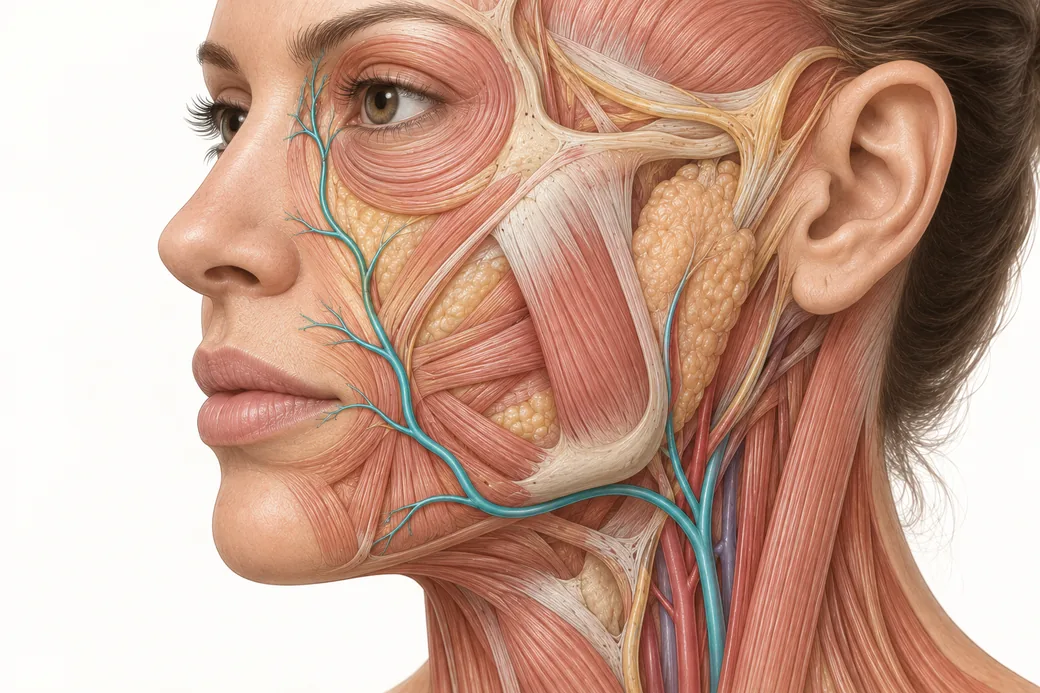 Clinical GuideAnastomoses: Why the Complication Doesn't Appear Where You InjectedFacial artery anastomoses explain why a lip treatment causes nasal skin changes. How arterial connections reframe injector triage and p
 Clinical GuideFiller Vascular Occlusion and Vision Loss: The Glabellar Crossroad, Step by StepHow filler vascular occlusion at the glabella causes vision loss — the retrograde embolic chain from the angular artery to the retina,
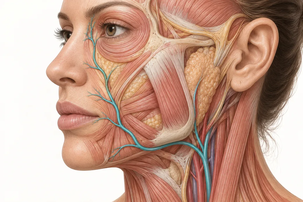 Clinical GuideFacial Artery Variability: Why Landmarks Are Probabilities, Not CertaintiesFacial artery variability in cadaver data — termination points, branch prevalence, tortuosity and depth, and what probabilistic anatomy
Train with Empire
This guide is clinical education. The technique behind it is taught hands-on, on live patients, with faculty beside you.
Explore Anatomical-Based Aesthetics Training →Disclaimer
This article reflects the clinical opinions and experience of Melissa Pulcini-Buttine, PA, an independent faculty member contributing to Empire Medical Training's curriculum. The views expressed are the author's own and do not necessarily represent those of Empire Medical Training.
It is professional education, not medical advice, and is no substitute for hands-on training or independent clinical judgment. Licensed clinicians remain responsible for their own patient selection, technique and outcomes, for verifying current product labelling, and for practising within their scope and applicable law. Empire Medical Training accepts no liability for reliance on this content.
 Claim Your Prize!