







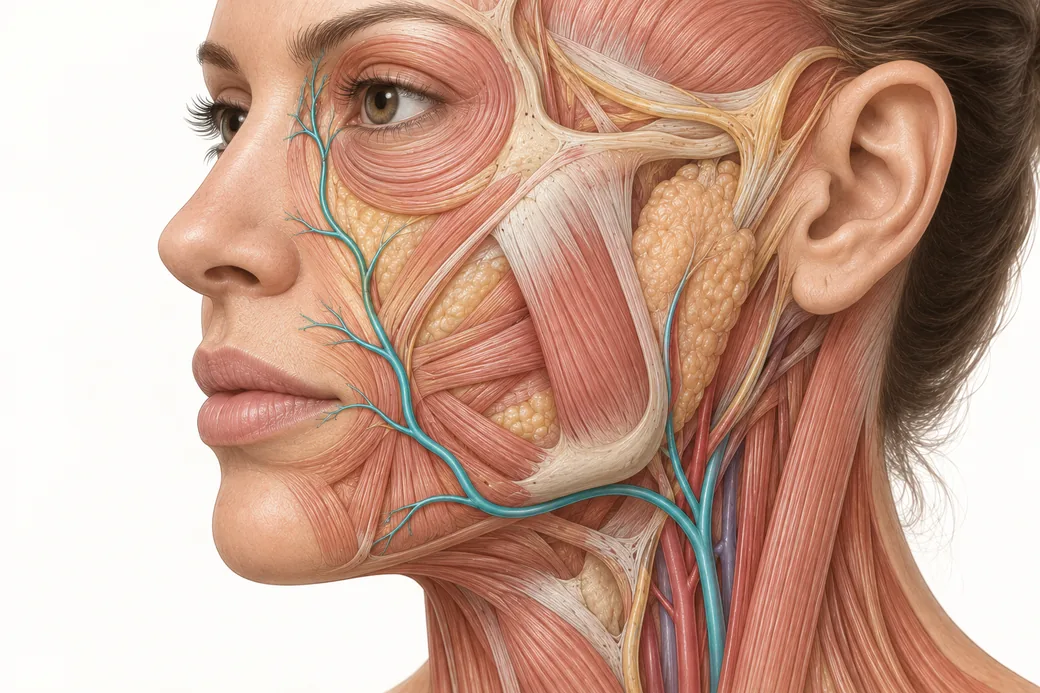
The call comes in two days after a lip treatment. The patient has dusky, mottled skin changes — on the nose. You did not inject the nose. You injected the lips.
The instinct, at that moment, is to doubt the history. Did she have something else done? Is this unrelated? Is she describing it accurately? Almost always, the history is fine and the anatomy is the explanation. Facial artery anastomoses — direct connections between arteries — mean that the territory you treated and the territory that failed are on the same circuit.
This is the most counterintuitive concept in facial vascular anatomy, and it is the one that most changes how an injector should triage a post-procedure phone call. Injectors are taught to think of an arterial tree: a trunk, branches, twigs, endpoints. The face is not a tree. It is a network, with cross-connections at multiple levels, redundant inflow, and — critically — the capacity for flow to move in a direction the textbook arrow does not show.
What an anastomosis actually is
An anastomosis is a communication between two vessels. Not a shared neighbourhood, not proximity — an actual continuous lumen connecting one arterial territory to another.
In most of the body, anastomoses are a feature. They are why a partial occlusion in one artery does not necessarily kill the tissue it feeds: a neighbour picks up the load. The face is one of the most richly anastomotic regions in the body precisely because it is a high-consequence, high-mobility, thin-skinned region that cannot afford ischaemia.
For a surgeon, this is protective. For an injector, it cuts both ways. The same connections that keep facial skin perfused under a narrowed pedicle also create reflux pathways — routes through which injected material can leave the territory you introduced it into. Work on the supratrochlear artery describes this explicitly: consistent anastomoses with the facial and angular systems provide cross-territorial inflow that maintains perfusion, and simultaneously create multiple reflux pathways during high-pressure filler injection (Merizaj et al., Cureus, 2025;17(9):e92563).
So the network property is not a defect in the anatomy. It is the anatomy working as designed, and you are the variable it did not evolve to accommodate.
The three crossroads worth knowing by heart
There are many anastomoses in the face. Three of them account for most of the "but I didn't inject there" phone calls in aesthetic practice.
The perioral–nasal crossroad
The superior labial artery runs medially through the upper lip. As it approaches the philtrum, it commonly gives off a columellar branch that ascends to the nasal base and septum. Published anatomical series put the superior labial artery as the source of the columellar supply in roughly 72.5% of specimens.
That is a direct, named arterial road from the upper lip to the nose. It is not subtle and it is not rare. Treat the lip or the philtral column, and the nasal base is anatomically downstream — or upstream, depending on direction of travel — on the same vessel.
This one crossroad explains the single most common surprise complication pattern in aesthetic injecting: lip in, nose out. The dedicated spoke on the perioral–nasal corridor in this cluster maps the substrate in detail.
The medial canthal crossroad
The facial artery, where it continues past the nasal ala, becomes the angular artery and terminates around the medial canthus. There it meets the dorsal nasal artery — which is not a facial artery branch at all. The dorsal nasal artery comes off the ophthalmic artery, a branch of the internal carotid.
So at the medial canthus, the external carotid system anastomoses with the internal carotid system. Two circulations that the diagram in your anatomy textbook draws as separate are, in this square centimetre, continuous.
This is the anastomosis with the highest ceiling of consequence, because the ophthalmic artery leads to the central retinal artery. The mechanistic chain from an angular artery embolus to retinal ischaemia is the subject of the glabellar crossroad spoke in this cluster.
The glabellar convergence
Just above the medial canthus, the supratrochlear and supraorbital arteries emerge from the orbit. Both are ophthalmic artery branches. Both are superficial — one series measured the superficial supratrochlear branch at around 4.2 mm depth in the central forehead, and the supratrochlear runs subcutaneously for much of its course (Phumyoo et al., Clinical Anatomy, 2020).
The glabella is therefore a small territory with a dense convergence of superficial vessels that connect downward to the angular artery and upward into the orbit. Small area, low margin for error, maximum connectivity. That combination is what makes it high-risk — not the label on the diagram.
Why the obstruction is usually not where the needle was
Here is the reframe that matters most clinically, and it is the one Empire's anatomy teaching pushes hardest.
When a complication presents, most injectors mentally mark the entry point and start there. But in an embolic event, the material does not stay at the point of entry. It is carried. Where it lodges is determined by flow direction, vessel calibre, bolus volume and branch geometry — not by where the needle tip was.
Material introduced under pressure into an artery can travel anterograde, with flow, into progressively smaller distal vessels until calibre stops it. Or it can travel retrograde, against flow, when the injection pressure at the needle tip exceeds systolic arterial pressure plus the frictional resistance of the column ahead of it. Once the plunger stops and pressure normalises, the material is then carried wherever flow takes it from that new position.
That second pathway is why an embolus can end up in a vessel proximal to — or in a completely different territory from — the one you entered. Most of the time, the occlusion is further up the circuit than the injection site. Anatomically, the obstruction sits somewhere on the connected territory, and the connected territory can be much larger than the treated one.
So the triage question is not "where did I inject?" It is:
- Which vessel does this entry point sit on or near?
- What does that vessel connect to, in both directions?
- Which of those territories is showing signs?
- Where, on that combined map, is an obstruction most likely to be sitting?
Answer those four and you are treating an anatomical problem. Skip to step four alone, and you are treating a puncture site.
Reframing the post-procedure phone call
Practically, this changes what you ask and what you look at.
Ask about the whole territory, not the treated site. A patient who calls about the treated area will describe the treated area. A patient with a nasal complication after lip treatment may not mention the nose because she doesn't think it's connected. Ask directly about the nasal tip and ala, the columella, the nasolabial region, the medial canthus and the forehead — the territories on the circuit. Ask about vision. Ask about pain that is disproportionate, not just visible change.
Look at the map, not the mark. Photograph and examine the connected territory. Blanching, mottling, dusky reticulated discoloration and delayed capillary refill in an untreated region after an injectable is a vascular finding, not a coincidence.
Do not let distance reassure you. "It's nowhere near where I injected" is the sentence that delays recognition. In a network, distance from the needle is weak evidence of anything.
Treat time as the scarce resource. Recognition speed is the variable you control after the fact. Everything about how to act on a suspected occlusion — the recognition criteria, the escalation pathway, dosing and the rescue protocol itself — sits in Empire's vascular occlusion material taught by Michelle Langston, and is deliberately not duplicated here. On the mechanism of dissolving hyaluronic acid specifically, see how filler is dissolved. For the broader picture of what patients report after lip treatment and how to separate expected from concerning, see common lip filler reactions and how to avoid them.
Two directions, two different clinical pictures
It is worth holding the distinction clearly, because it predicts what you will see.
Anterograde embolisation produces signs in the distal territory of the vessel entered. It tends to present as skin-level ischaemia in a recognisable vascular distribution: the ala and nasal tip for the lateral nasal artery, the lip for a labial artery, the nasolabial and cheek skin for a facial artery branch.
Retrograde embolisation produces signs that can appear in a territory the entered vessel connects to rather than feeds. Its worst expression is ocular — material driven back through the angular or supratrochlear artery into the ophthalmic circulation. The published case literature bears out that the combination is common rather than exceptional: in the largest review of filler-associated vision loss, skin changes accompanied the visual event in 73.2% of cases (Doyon et al., Aesthetic Surgery Journal, 2024;44(10):1091–1104). Skin and eye are frequently the same event, seen from two ends of the network.
That statistic has a practical corollary. Skin findings after a facial injectable are not only a skin problem. They are a marker that material went where it should not have gone, and they warrant a question about vision every time.
Where the reasoning has limits
Evidence honesty matters more in anatomy than anywhere else, so two caveats.
First, anastomoses are as variable as the vessels they join. The columellar branch is present bilaterally in only a minority of specimens in some series, unilaterally in most, and absent in a meaningful fraction. The angular artery itself is present in roughly a third to three-quarters of hemifaces depending on the series. You cannot assume a specific connection exists in a specific patient — you can only assume that some connections exist, which is why the safety margin is the answer rather than the map alone.
Second, the direction and destination of an embolus are not predictable at the bedside. The anatomical reasoning above tells you where to look. It does not tell you where the material is. Nobody can tell you that from the outside. The reasoning narrows the search; it does not replace examination of the entire connected territory.
What the network concept genuinely buys you is earlier recognition and a wider field of view. That is not a small thing. In a condition where outcome is dominated by time to intervention, a clinician who checks the nose after a lip treatment is operating on a different timeline from one who doesn't.
Learning connections you can see
Anastomoses are the part of facial anatomy that reads as abstract on a page and becomes obvious the moment you see one. You can be told that the superior labial artery talks to the nasal base. Following that vessel yourself, in tissue, is what makes you check the nose on a Tuesday afternoon phone call three years later.
That is what Empire's Special Anatomical Cadaver Aesthetics Training is built to deliver, and why the anatomy comes before the technique in Complete Dermal Filler Training.
These anatomical relationships reflect Melissa Pulcini-Buttine's teaching in Empire Medical Training's hands-on curriculum, supported by the published literature cited above. This article is educational and is not a substitute for supervised training or for your practice's vascular complication protocol.
Melissa Pulcini-Buttine, PA — physician assistant of two decades; professor of anatomy and physiology for ~14 years; faculty member, Empire Medical Training; founder of an aesthetics practice in Greenwich, Connecticut.
Related guides in this cluster
Part of Facial Vascular Anatomy for Injectors.
 Clinical GuideFiller Vascular Occlusion and Vision Loss: The Glabellar Crossroad, Step by StepHow filler vascular occlusion at the glabella causes vision loss — the retrograde embolic chain from the angular artery to the retina,
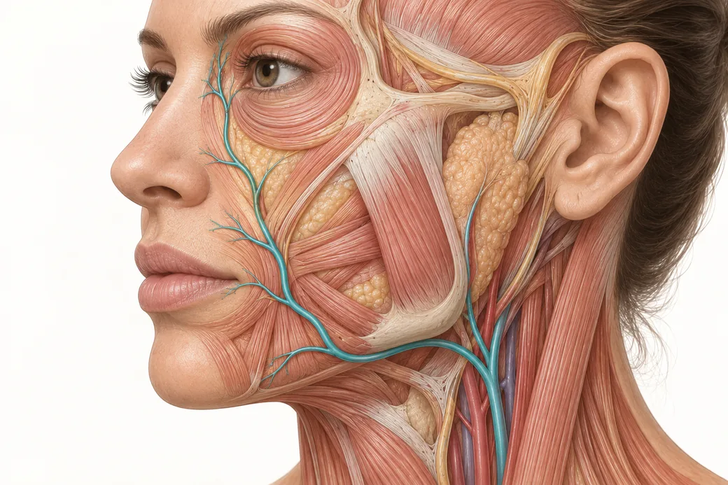 Clinical GuideFacial Artery Variability: Why Landmarks Are Probabilities, Not CertaintiesFacial artery variability in cadaver data — termination points, branch prevalence, tortuosity and depth, and what probabilistic anatomy
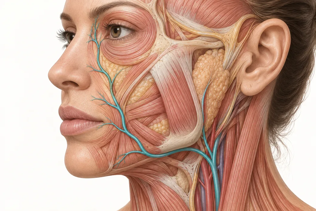 Clinical GuideThinking in Layers: Depth as the Second Axis of the Vascular MapFacial layers explained for injectors — the five-layer model, which layer each facial artery occupies, and why a depth in millimetres m
Train with Empire
This guide is clinical education. The technique behind it is taught hands-on, on live patients, with faculty beside you.
Explore Anatomical-Based Aesthetics Training →Disclaimer
This article reflects the clinical opinions and experience of Melissa Pulcini-Buttine, PA, an independent faculty member contributing to Empire Medical Training's curriculum. The views expressed are the author's own and do not necessarily represent those of Empire Medical Training.
It is professional education, not medical advice, and is no substitute for hands-on training or independent clinical judgment. Licensed clinicians remain responsible for their own patient selection, technique and outcomes, for verifying current product labelling, and for practising within their scope and applicable law. Empire Medical Training accepts no liability for reliance on this content.
 Claim Your Prize!