







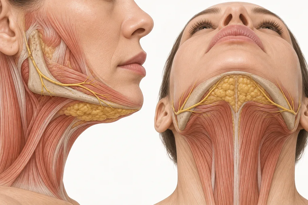
The submental pinch test is the cheapest diagnostic in aesthetic medicine and the one that most often gets skipped. It takes four seconds, costs nothing, and is the difference between a patient who thanks you at twelve weeks and a patient who tells you their neck looks worse. A cytolytic fat-reduction drug can only act on fat. If what you are looking at is skin, you can inject perfectly and still fail.
My rule is the shortest one I teach: treat what you can pinch.
Maritza Mejia, FNP — family nurse practitioner; faculty member, Empire Medical Training; founder of Long Island Beauty Bar, New York.
Why a pinch is a diagnostic and not a gesture
A submental convexity has a limited number of possible causes, and only one of them is treatable with deoxycholic acid.
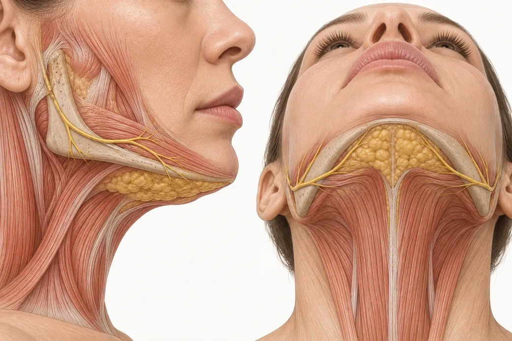
Larson and colleagues weighed every fat compartment in ten fresh cadaver necks (Aesthetic Surgery Journal, 2014; PMID 24633742) and found that supraplatysmal fat — the compartment between skin and platysma — accounted for on average 44.7% of the fat in the neck. Subplatysmal fat, deep to the muscle, accounted for 30.7%. The submandibular gland accounted for 24.5%. "Very deep" fat, deep to the anterior digastrics, was under 1%.
Read that as a clinician holding a vial. Injectable deoxycholic acid is labelled for injection into pre-platysmal subcutaneous fat. That means the drug can address, at most, something under half of the fat volume in a neck — and none of the gland, which is a quarter of it. Everything that makes a neck look full and is not supraplatysmal fat is outside the drug's reach.
The pinch is how you find out which compartment you are looking at, because the pinch physically isolates the supraplatysmal layer. Skin and the fat superficial to platysma lift away between your fingers. Subplatysmal fat, the gland, the digastrics and a low-lying hyoid do not.
How Maritza performs it
The manoeuvre I teach is deliberately unglamorous.
Position the patient relaxed and upright. Not lying down, not chin-up, not clenching. A relaxed head-neutral seated position is the reference state. Everything else in the examination sequence is a departure from it.
Pinch and fold the tissue between your thumb and your index finger. One hand. The thumb sits below the mandibular border, the index finger comes up from beneath, and the two close over the submental tissue in the midline. You are lifting, not squeezing.
Assess the thickness of the fold, not the length of it. A long fold of thin tissue and a short fold of thick tissue tell you opposite things. The number you want is how deep the pinch is between your fingertips.
Repeat it across the area, not once in the middle. Submental fat is not uniform. I pinch midline, then either side of midline, then out towards the lateral submental region and the submandibular extension. A pinch that is generous centrally and empty laterally defines the treatable footprint, and that footprint is what the mapping grid will later cover.
Then palpate without pinching. After the pinch, run flat fingers over the same territory to feel for anything firm, lobulated or discrete — gland, node, scar, thyroid enlargement. The pinch tells you how much soft tissue is mobile; the palpation tells you what else is in the field.
Reading the fold: Maritza's thickness bands
Here are the figures I use at the chairside:
| Pinch thickness | What it most likely means | What I do |
|---|---|---|
| Roughly 2–3 cm of soft, compressible tissue | Fat is most likely the dominant cause of the convexity | Proceed to full assessment and mapping |
| Less than about 1 cm of tissue | The convexity is more likely loose skin than fat | Stop. This is a laxity problem, not a fat problem |
| Between those, or firm rather than soft | Mixed picture, or something other than fat in the fold | Re-examine dynamically before deciding |
These are the thresholds I work to at the chairside, and they sit inside a full assessment rather than replacing one. They give you a way to put a number on what your fingers are already telling you, which matters when you are building a record you can defend and when you are comparing session one against session three.
They also line up with how patient selection is described in this drug's own literature: the jowl-fat study by Shridharani and colleagues (Plastic and Reconstructive Surgery, 2020; PMID 32221205) enrolled patients with "pinchable fat" and "relatively minimal skin laxity" — the same two findings, read the same way. Severity itself is scored separately, on the Clinician-Reported and Patient-Reported Submental Fat Rating Scales used as endpoints in the pivotal trials, and those scales are what you document alongside the pinch.
The principle underneath all of it is in the label: "Prior to each treatment session, palpate the submental area to ensure sufficient submental fat."
These figures reflect Maritza Mejia's clinical practice as taught in Empire Medical Training's hands-on curriculum. Technique is learned under supervision; this article is educational and is not a substitute for training.
The four things a pinch can tell you apart
Fat
Soft, compressible, uniformly thick across a region rather than in a discrete lump, lifts freely from the underlying muscle, and — this is the tell — the fold stays substantial when the patient's neck is relaxed. Treatable, if the rest of the assessment agrees.
Skin laxity
The fold is thin, the skin is the fold. When you release it, it settles slowly or creases. The convexity is a drape problem: there is more envelope than content. Reducing content in a neck like this does not improve the drape and may expose it. Deoxycholic acid is the wrong tool; the conversation is about tightening or lifting modalities, and thread lifting for a double chin is one of the alternatives worth understanding before you refer.
Submandibular gland
Firm, often lobulated, sits below and behind the mandibular angle rather than in the midline, and — crucially — does not lift into a pinch. It is a fixed structure, not a mobile layer. A ptotic submandibular gland is a common reason a neck looks full after successful fat reduction, and the patient will read that as your failure if you did not name it in advance. The label explicitly instructs that the drug should not be injected into or in close proximity (1–1.5 cm) to salivary glands.
Lymph node
Discrete, mobile, round, usually singular, and it rolls under your finger rather than folding. A node in the submental triangle is a referral question before it is an aesthetic one.
The decision rule
This is the gate. I do not open a vial until all four of these are true.
- There is a pinchable fold of soft tissue across the intended treatment footprint — not just at one point in the midline.
- The fold is fat-dominant, not skin-dominant, on the thickness assessment above.
- Nothing firm, lobulated, discrete or fixed is in the field, or if it is, it has been identified and excluded from the map.
- The patient's dominant stated concern matches what the pinch found. A patient who says "I hate the loose skin" and has 2.5 cm of pinchable fat is still a patient whose expectation will not be met.
If any of those four fails, the answer is not a smaller dose. The answer is a different plan.
Why a pinch is not the whole examination
The pinch is a static test performed in one position, and it is deliberately narrow. It answers one question — is there treatable fat here — and it answers it well. It does not tell you about platysmal bands, which only declare themselves on animation. It does not tell you about redundancy, which only declares itself on flexion. It does not tell you whether a full-looking neck is a low hyoid or a ptotic gland, which extension and swallowing will reveal.
That is why the pinch sits inside a longer sequence — resting, smiling, flexion, extension, palpation, photography — rather than replacing it. Use the pinch as the go/no-go gate on fat. Use the dynamic examination to decide whether fat reduction alone will produce the result the patient is imagining.
The pinch also has a second life during the procedure itself. The label's technique instruction has the patient tense the platysma while the operator pinches the submental fat and injects perpendicular to the skin into the pre-platysmal fat. The same manoeuvre that selected the patient is what keeps the needle in the right compartment once you are working. And the label instructs palpation before each treatment session, not just the first — which makes the pinch a retreatment decision as much as an initial one.
What changes at the chairside
- Pinch before you quote. Deciding candidacy after a patient has been given a price is a bad position to negotiate from.
- Pinch the whole footprint, not the midline. The footprint you can pinch is the footprint you can map.
- Write the pinch findings down. "Pinchable soft tissue midline and bilaterally; no firm or discrete structures palpable; laxity mild" is a defensible record. "Suitable for treatment" is not.
- Say the word "no" out loud when it applies. The patients who damage a practice are the ones who were treated with the wrong tool, competently.
- Re-pinch before every subsequent session. Sufficient fat at session one does not mean sufficient fat at session three, and the label requires the check.
Injectors who want to practise assessment, mapping and injection on live patients under faculty supervision can find that in Empire's Kybella and Mesotherapy Training. If the necks you are seeing are mixed fat-and-laxity presentations, the combination-therapy approach taught in the Neck & Hands Rejuvenation Master Course is the more relevant training.
Related guides in this cluster
Part of Submental and Lower Face Contouring.
 Clinical GuideHow Deoxycholic Acid Works: Adipocytolysis, Inflammatory Clearance, and Why Swelling Is the TreatmentHow deoxycholic acid causes adipocytolysis, why the inflammatory swelling that follows is the clearance mechanism, and what that change
 Clinical GuideSubmental Anatomy for Deoxycholic Acid: Compartments, the Submandibular Extension and the Marginal Mandibular NerveThe safety anatomy of the neck for injectors — preplatysmal versus subplatysmal fat, the submandibular extension, and where the margina
 Clinical GuideDynamic Neck Assessment: Resting, Smiling, Flexion and ExtensionA four-position neck examination for injectors — what rest, animation, flexion and extension each reveal that a static pinch cannot, an
Train with Empire
This guide is clinical education. The technique behind it is taught hands-on, on live patients, with faculty beside you.
Explore Complete Dermal Filler Training →Disclaimer
This article reflects the clinical opinions and experience of Maritza Mejia, FNP, an independent faculty member contributing to Empire Medical Training's curriculum. The views expressed are the author's own and do not necessarily represent those of Empire Medical Training.
It is professional education, not medical advice, and is no substitute for hands-on training or independent clinical judgment. Licensed clinicians remain responsible for their own patient selection, technique and outcomes, for verifying current product labelling, and for practising within their scope and applicable law. Empire Medical Training accepts no liability for reliance on this content.
 Claim Your Prize!