







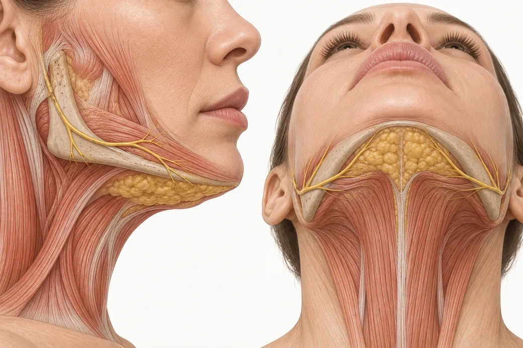
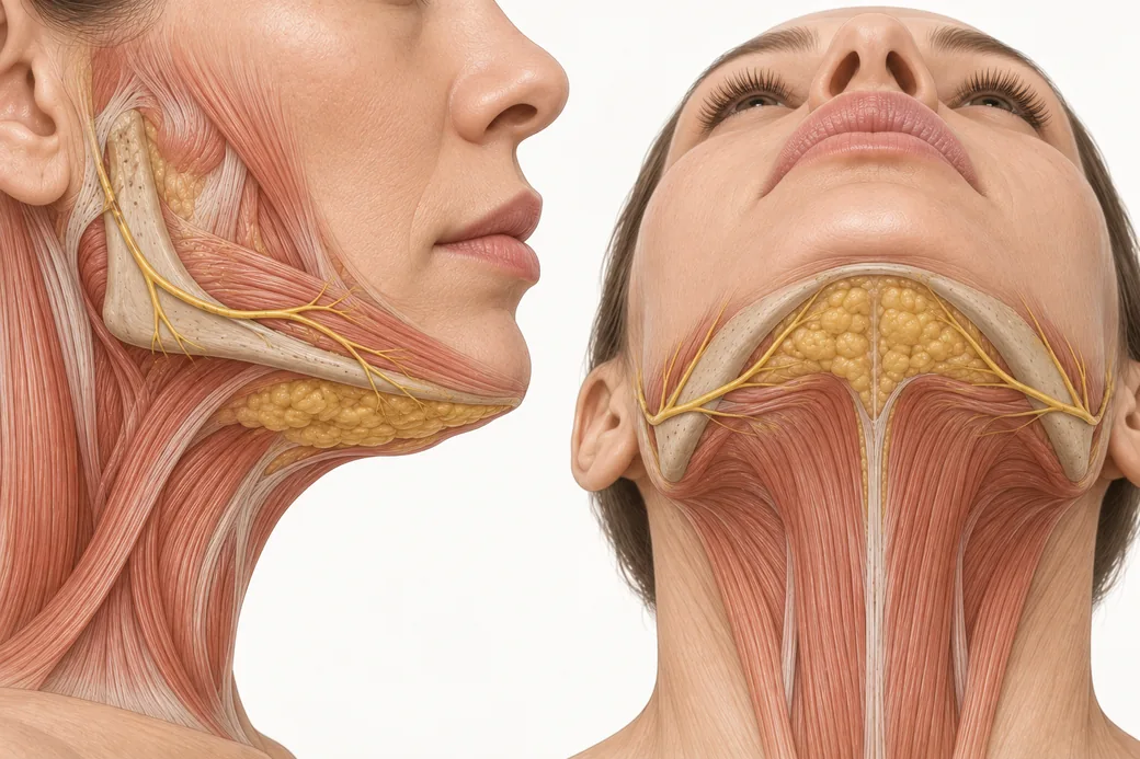
Aesthetic injectors carry a detailed map of the face and a blank space below the jawline. The marginal mandibular nerve is the single most consequential structure in that blank space, and a submental fat-reduction treatment puts a needle into its territory fifty times in one sitting. Anatomy decides the plan. This is the regional anatomy that decides it.
Everything here is oriented around one practical question: when you place 0.2 mL of a cytolytic drug into a neck, what is above it, what is below it, what is beside it, and what happens if you miss.
Maritza Mejia, FNP — family nurse practitioner; faculty member, Empire Medical Training; founder of Long Island Beauty Bar, New York.
The layered neck, superficial to deep
Under the submental skin, in order:
- Skin.
- Supraplatysmal (pre-platysmal) subcutaneous fat. This is the target compartment. It is the only compartment a labelled deoxycholic acid injection is meant to enter.
- Platysma. A broad, thin sheet of muscle. Its medial edges are what cord into visible bands.
- Subplatysmal fat. Deep to the muscle, sitting between the medial edges of the anterior digastric muscles in the midline.
- Anterior digastric muscles, and the submandibular glands laterally.
- "Very deep" fat, deep to the digastrics and gland, adherent to the strap muscles.
- The mylohyoid and the floor of the mouth.
Larson and colleagues dissected and weighed these compartments in ten fresh cadaver heads (Aesthetic Surgery Journal, 2014; PMID 24633742). The proportions are the most clinically useful numbers in this entire article:
| Compartment | Share of neck fat volume |
|---|---|
| Supraplatysmal (the treatable one) | 44.7% |
| Subplatysmal | 30.7% |
| Submandibular gland | 24.5% |
| Very deep (deep to digastrics) | <1% |
Both the supraplatysmal and subplatysmal compartments were further divided into suprahyoid and infrahyoid portions. Gassman and colleagues (Plastic and Reconstructive Surgery, 2017; PMID 28841605) made the related point that the microscopic septal architecture that defines discrete facial fat compartments is present in the neck as well — the neck is compartmentalised tissue, not an undifferentiated layer.
Read those percentages as a dosing ceiling. An injector working in the correct plane has access to a compartment that is, on average, under half the fat in the neck. The remaining 55% is either behind a muscle or is not fat at all. No volume of drug placed correctly reaches it, and any attempt to reach it means leaving the safe plane. This is why some technically perfect treatments produce a modest contour change: the fat you could reach was never the fat that was dominating the silhouette.
Why the plane is the primary safety mechanism
Two separate protections keep this drug from injuring structures it should not touch, and they are not equally strong.
The weaker protection is tissue selectivity. Thuangtong and colleagues (Dermatologic Surgery, 2010; PMID 20482723) showed that deoxycholate lyses every cell type in vitro, but that albumin and protein-rich tissue reduce its lytic capacity — leaving protein-poor adipose tissue uniquely vulnerable. That buffering is real, and it is why a stray molecule diffusing into muscle does less than the same molecule in fat. It is not a guarantee, and it does nothing about the inflammatory field that follows lysis.
The stronger protection is depth. Every structure you are trying to avoid — platysma, digastrics, submandibular gland, the vessels of the submental triangle, and the marginal mandibular nerve — lies deep to the platysma or deep to the superficial fascial plane. The pre-platysmal subcutaneous fat is, anatomically, a separate floor of the building. Walker's Phase 1 histology study (Dermatologic Surgery, 2020; PMID 30883481) found that across every concentration, volume and spacing paradigm tested, the histological changes were confined to the subcutaneous layer. Correct depth contains the injury.
This is why the label's technique instruction reads the way it does: have the patient tense the platysma, pinch the submental fat, and inject perpendicular to the skin into the pre-platysmal fat with a 30-gauge needle. Tensing the platysma firms the floor. Pinching lifts the target away from it. The two manoeuvres together convert an anatomical plane into something you can feel.
The marginal mandibular nerve: what the label says
The label gives a geometric exclusion zone:
Do not inject above the inferior border of the mandible. Do not inject within a region defined by a 1–1.5 cm line below the inferior border (from the angle of the mandible to the mentum).
That zone exists because marginal mandibular nerve injury was observed in the pivotal trials. Nerve injury occurred in 4% of treated subjects versus under 1% on placebo. It presents as an asymmetric smile or facial muscle weakness on the affected side — the nerve supplies the depressor anguli oris, depressor labii inferioris and mentalis, so the affected corner of the mouth fails to depress and the smile pulls unevenly.
The reassuring part: all marginal mandibular nerve injuries reported from the trials resolved spontaneously, with a range of 1 to 298 days and a median of 44 days. The sobering part: a median of 44 days is six weeks of a patient looking in the mirror at an asymmetric smile that you caused, and a range extending to ten months.
The label also gives a proximity rule for the other structures in the region: the drug should not be injected into or in close proximity (1–1.5 cm) to salivary glands, lymph nodes and muscles.
The marginal mandibular nerve: what the cadavers say
Here is the part that is not on the label and that every injector working in this region should know.
The 1–1.5 cm exclusion zone is a practical compromise, not a guarantee that the nerve lies outside it. Cadaveric series disagree with one another — partly because they measure from different reference points and in different neck positions — but they converge on one message: the nerve's course is variable, and in a minority of people the lowest branch runs further inferiorly than the rule of thumb predicts.
- Sindel et al. (British Journal of Oral and Maxillofacial Surgery, 2021; PMID 33483156) dissected twelve fresh cadavers, contouring the mandibular base at 5 mm intervals. The highest levels of the nerve were 6.9 mm and 6.5 mm above the mandibular base; the lowest were 4 mm and 3 mm below it.
- Anthony et al. (Patient Safety in Surgery, 2018; PMID 30159033) measured the maximum perpendicular distance from the lowest ramus of the marginal mandibular branch to the inferior border of the mandible in 22 cadavers: mean 7.12 ± 2.97 mm, maximum recorded 17.65 mm.
- Davies et al. (Clinical Anatomy, 2016; PMID 26096443) compared the commonly taught "2 cm below the mandible" and "two finger breadths" landmarks against 3D-digitised nerve courses in 31 specimens. In 7 of 31 specimens the most inferior branch coursed below the 2 cm line. The two-finger-breadth line, by contrast, was never crossed — but its position varied from 25 to 51 mm below the mandibular border depending on whose fingers were used, which makes it useless as a standard.
- Karapinar et al. (Saudi Medical Journal, 2013; PMID 23552588) found the nerve presented as a single branch in 36.4% of hemifaces and two branches in 63.6%, running lateral to the facial artery in 97.7%.
Synthesise those and the honest teaching point is this: the nerve is usually above or just below the mandibular border, but it can run a centimetre or more below it, and a geometric line does not know which patient is in front of you. The exclusion zone reduces risk. The injection plane is what actually protects the nerve, because the marginal mandibular nerve runs deep to platysma along most of its aesthetic course. A needle held in pre-platysmal fat is in a different compartment from the nerve regardless of where the nerve happens to run.
A related structural finding: Yalçın and colleagues (Aesthetic Surgery Journal, 2026; PMID 42056850) reassessed the mandibular septum and found the marginal mandibular nerve approximately 8.4–8.7 mm inferior to the septum at the gonial angle, with the nerve crossing the mandible at the junction of the mandibular septum and the platysma-mandibular ligament. The septum and ligament act as a barrier limiting inferior migration of jowl fat into the neck — which is also why jowl fat and submental fat behave as distinct problems.
The submandibular extension and the lateral compartment
Patients rarely present with fullness confined to a tidy midline triangle. The fat that bothers them frequently extends laterally and posteriorly under the mandibular body — what I teach as the submandibular extension of the treatment area.
This is the most dangerous real estate in the region, and for four separate reasons stacked in the same few square centimetres:
- The marginal mandibular nerve is closest to the surface and closest to your grid here, near the mandibular border and the angle.
- The submandibular gland sits here. Firm, lobulated, and a structure the label says to stay 1–1.5 cm away from. In a neck with gland ptosis the gland is lower than you expect, which narrows your working corridor further.
- The facial artery and vein cross the mandibular border at the antegonial notch, anterior to the masseter. The marginal mandibular branch runs lateral (superficial) to the facial artery in the large majority of specimens.
- Submental and submandibular lymph nodes sit in the field, and the label instructs staying 1–1.5 cm away from lymph nodes as well.
The practical rule I use when mapping: the exclusion zones get marked first, before any treatment grid is drawn. The mandibular border is marked. The 1–1.5 cm inferior line from the angle to the mentum is marked. Anything firm, lobulated or discrete that palpation found is marked. Only then do I lay a treatment grid inside what is left. The grid never defines the boundary; the anatomy defines the boundary and the grid fills it.
Dysphagia, and what it tells you about diffusion
Difficulty swallowing occurred in 2% of treated subjects in the pivotal trials versus under 1% on placebo, in the setting of administration-site reactions. Cases resolved spontaneously with a range of 1 to 81 days and a median of 3 days.
The mechanism is instructive. Most of those cases are not a drug injury to a swallowing structure — they are the consequence of substantial oedema in a confined anterior neck. A tissue plane that swells presses on what is beneath it. That is also why the label advises against use in patients with pre-existing dysphagia: you are adding a temporary mass effect to a system that already has less reserve.
It is also a reminder that the inflammatory field is not confined by the same boundaries as the drug. The drug stays in the subcutaneous layer; the swelling it produces is regional.
What good anatomical practice looks like in this region
- Mark exclusion zones before treatment zones. Danger first, target second.
- Keep the patient's platysma tensed and keep the tissue pinched during injection. These are not ceremonial; they are what converts an anatomical plane into a tactile one.
- Inject perpendicular into the middle of the pinched subcutaneous layer. Too superficial risks the dermis and ulceration or necrosis; too deep risks everything in this article.
- Consider the lidocaine check. Shridharani and colleagues (Aesthetic Surgery Journal Open Forum, 2024; PMID 39247122) describe injecting lidocaine and watching for smile asymmetry as an indicator of marginal mandibular nerve proximity before committing the treatment agent — a reversible test with an irreversible alternative.
- Document a pre-treatment smile. If a patient develops asymmetry, the only way to know whether it is new is a baseline photograph of them smiling. Standardised photography is the medicolegal spine of this treatment, and it is worth following your practice's canonical clinical photography protocol rather than improvising views.
- Know which structures your training has and has not covered. The face has a mature danger-zone literature for injectors. The neck does not, and the difference in confidence between the two regions is not matched by a difference in risk.
These figures reflect Maritza Mejia's clinical practice as taught in Empire Medical Training's hands-on curriculum, together with the current FDA prescribing information and the published anatomical literature cited above. Technique is learned under supervision; this article is educational and is not a substitute for training.
Injectors whose gap is regional anatomy rather than product knowledge should start with Anatomical Based Aesthetics Training or, for dissection-based work, Special Anatomical Cadaver Aesthetics Training. Submental mapping and injection on live patients under faculty supervision is covered in Kybella and Mesotherapy Training, and the regional combination approach in the Neck & Hands Rejuvenation Master Course. For neurotoxin site selection in the lower face, see Botox injection sites.
Related guides in this cluster
Part of Submental and Lower Face Contouring.
 Clinical GuideHow Deoxycholic Acid Works: Adipocytolysis, Inflammatory Clearance, and Why Swelling Is the TreatmentHow deoxycholic acid causes adipocytolysis, why the inflammatory swelling that follows is the clearance mechanism, and what that change
 Clinical GuideThe Submental Pinch Test: Distinguishing Fat from Skin Laxity Before You Open a VialThe submental pinch test separates treatable fat from skin laxity. Maritza Mejia's thumb-and-index technique, thickness bands, and the
 Clinical GuideDynamic Neck Assessment: Resting, Smiling, Flexion and ExtensionA four-position neck examination for injectors — what rest, animation, flexion and extension each reveal that a static pinch cannot, an
Train with Empire
This guide is clinical education. The technique behind it is taught hands-on, on live patients, with faculty beside you.
Explore Complete Dermal Filler Training →Disclaimer
This article reflects the clinical opinions and experience of Maritza Mejia, FNP, an independent faculty member contributing to Empire Medical Training's curriculum. The views expressed are the author's own and do not necessarily represent those of Empire Medical Training.
It is professional education, not medical advice, and is no substitute for hands-on training or independent clinical judgment. Licensed clinicians remain responsible for their own patient selection, technique and outcomes, for verifying current product labelling, and for practising within their scope and applicable law. Empire Medical Training accepts no liability for reliance on this content.
 Claim Your Prize!