








Meralgia paresthetica is burning, numbness and altered sensation over the outer thigh, caused by compression of the lateral femoral cutaneous nerve as it passes near the groin. It is one of the few nerve entrapments where the diagnosis, the treatment and the reassurance all follow from a single fact about the nerve.
That fact, as Dr. Gisele J. Girault puts it, is that “the lateral femoral cutaneous nerve is only a sensory nerve. There are no motor fibers in it.” Nothing weakens. What the patient gets instead is a large, uncomfortable territory — “a fairly large area that patients will have a lot of burning and sort of dysaesthetic sensations” — which she describes as quite distressing even though nothing is at risk.
This reference covers the nerve’s territory, the situations that compress it, how it is separated from a lumbar radiculopathy, and the block. It sits under our clinical reference to joint and extremity injection.
Where the nerve runs and what it supplies
Girault describes the course simply: the lateral femoral cutaneous nerve is purely sensory, comes down through the groin, and goes to the lateral thigh.
Dr. Robert Stall maps the territory in more detail, and the detail is what lets a clinician match a patient’s drawing of their symptoms to this nerve rather than another. The nerve gives rise to two branches:
- The anterior branch supplies the skin over the anterolateral thigh and over the top of the iliotibial band down to the knee
- The posterior branch innervates the skin over the greater trochanter, extending distally to the mid-thigh, and sometimes extending posteriorly into part of the gluteal region
That posterior branch explains presentations that otherwise look wrong for the diagnosis — symptoms over the side of the hip or edging into the buttock are still within this nerve’s field, and do not require a second explanation.
Both faculty locate the nerve against the same landmark: the anterior superior iliac spine. Stall identifies the most proximal site to palpate for a treatment point as just inferior to the ASIS, where the nerve passes and courses laterally and distally. Girault palpates the ASIS and moves approximately two centimetres medial to it.
What compresses it
Girault’s three populations are all about sustained external pressure at the groin, and they are specific enough to be diagnostic on their own.
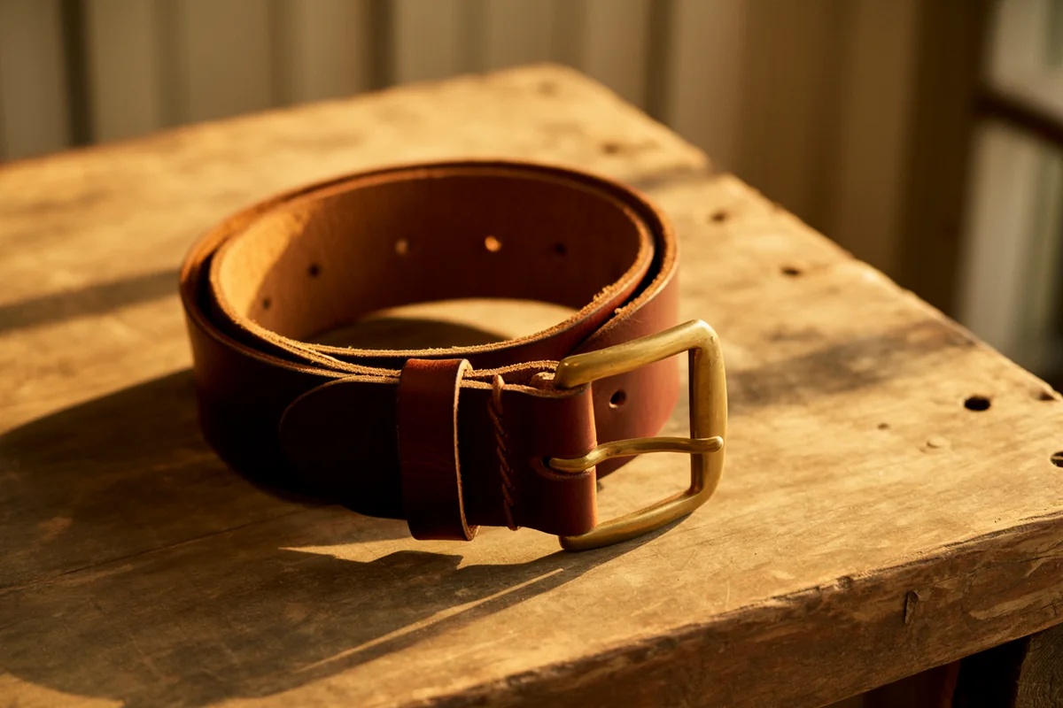
Load-bearing belts. She sees it predominantly in men carrying a tool belt — a police officer’s duty belt is her example — where the belt rests directly on the nerve as it passes through the groin.
Prolonged lithotomy position. In women who have been in lithotomy position for a prolonged period during a delivery.
A large pannus. In patients with an overhanging abdomen, where the weight rests on the nerve and injures it.
The pattern generalises. Anything that presses steadily on the region just inside the ASIS — a tight waistband, a heavy belt, a car seatbelt in an unusual position, body armour, sustained positioning during surgery — will do the same thing, which is why the history is often more informative than any test.
Separating it from a back problem
Outer thigh pain is commonly attributed to the lumbar spine, and the absence of motor involvement is what settles it.
A lumbar radiculopathy involves a nerve root carrying both motor and sensory fibres, so weakness, reflex change or symptoms extending below the knee in a dermatomal pattern belong to the root rather than to this nerve. Meralgia paresthetica produces a patch of abnormal sensation on the thigh and nothing else.
The other neighbours are the adjacent cutaneous nerves of the groin, and Girault’s method for sorting them is worth copying. When a patient presents with groin or anterior thigh pain, she takes out an anatomy book, looks at where they are pointing, and works out which nerve is involved — the anterior femoral cutaneous, the lateral femoral cutaneous, the genitofemoral or the ilioinguinal. Most of those presentations, she notes, follow abdominal surgery: a caesarean section, a hysterectomy, or an inguinal or femoral hernia repair, with the nerve caught in scar.
Where a nerve genuinely needs to be shown to be failing rather than merely irritated, electrodiagnostic testing answers that question — though a purely sensory nerve in a patch of thigh is a harder study than a mixed nerve in a limb.
The block
This is among the more forgiving injections in the lower limb, and Girault says so explicitly.
The nerve sits in fat, very superficially — on ultrasound she describes it perhaps half a centimetre from the surface in a thin patient, medial and superficial to the sartorius muscle and the tensor fasciae latae, with no blood vessels around it. The femoral nerve, artery and vein are some distance away, and she palpates for the femoral pulse each time to confirm she is clear of them.
Her technique uses the ASIS as the landmark, palpating it and moving about two centimetres medial. The injection is delivered in a fan-like manner, with roughly three to five cc of local anaesthetic plus steroid for a total of four to five cc. Her assessment of the risk is unusually plain: there is “not really anything that you can hit that’s going to do any significant damage in this area.”
She places it among the cutaneous nerve blocks of the groin — alongside the anterior femoral cutaneous, iliohypogastric and ilioinguinal nerves — that are reasonable when first starting out, while advising that the genitofemoral nerve and its branches are more advanced and better left alone.
Relief can be substantial where this nerve is genuinely the source. It is also diagnostic: a block that abolishes the symptoms confirms the territory, and one that does nothing argues the problem is elsewhere.
Whether to use ultrasound
Girault performs these blocks on landmarks and considers the region safe enough to do so, while noting that the nerve images clearly.
Guidance earns its place in the patients where palpation is least reliable — and those are precisely the patients most likely to have the condition, since an overhanging pannus both causes the compression and obscures the ASIS. The general case for guidance is set out in our musculoskeletal ultrasound reference.
Removing the pressure
The injection treats the irritated nerve. It does not remove the belt.
Where the cause is identifiable — and in this diagnosis it usually is — addressing it is what determines whether the relief lasts. Redistributing the load of a duty belt or tool belt, changing waistband height, and weight reduction where a pannus is responsible all act on the mechanism rather than the symptom. Where the compression was a one-off, such as prolonged positioning during a delivery or an operation, symptoms often settle on their own and the block serves to make the interval tolerable.
For clinicians
Cutaneous nerve blocks of the groin are landmark techniques learned on live anatomy with a proctor present, and the ones near the femoral vessels demand that orientation be confirmed before every injection. Empire teaches the lower extremity and groin blocks across the pain management academy.
Learn blocks with your hands, not from a page
Empire’s Pain Management Training (THE Pain Show) is accredited for 25.25 AMA PRA Category 1 Credits™, jointly provided by AKH, Inc, and Empire Medical Training. For narrower peripheral work, Joint, Extremity and Non-Spinal Injection Training carries 6.75 credits for the complete in-person hybrid program, and Advanced Musculoskeletal Ultrasound Guided Injections builds the guidance skills above.
Explore THE Pain Show Claim Your Prize!