







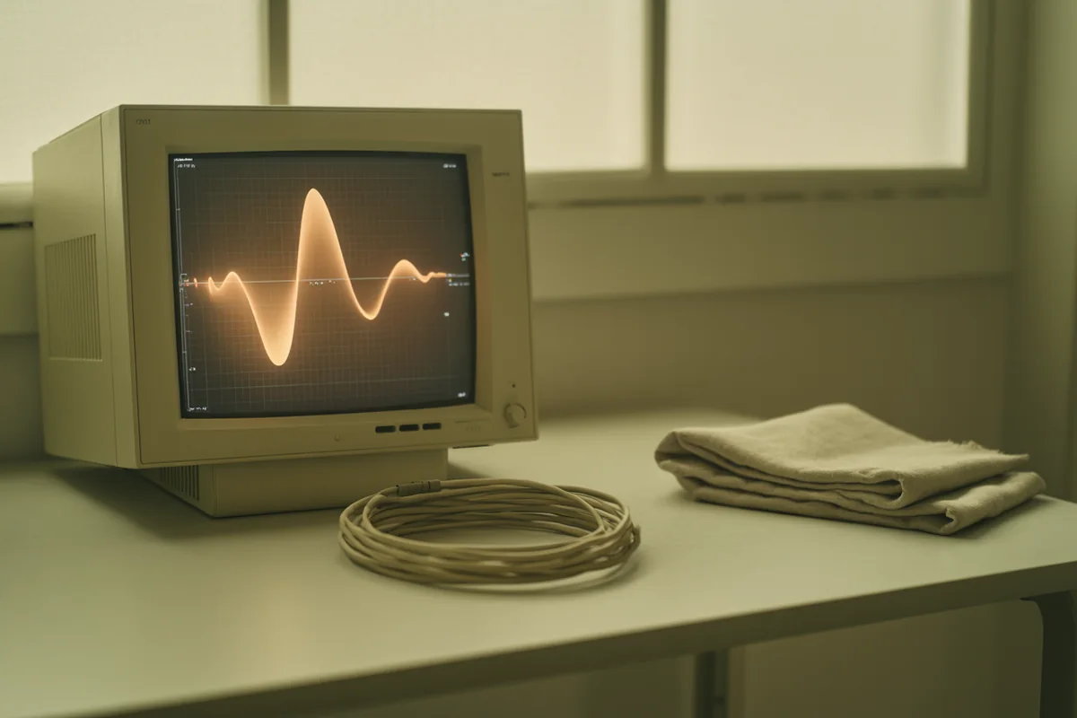

An electrodiagnostic study is two tests performed in one appointment. The nerve conduction study measures how well electrical signals travel along a nerve. The needle electromyogram listens to the electrical behaviour of the muscle that nerve supplies. Together they answer a question imaging cannot: not what a structure looks like, but whether the nerve is actually working.
This reference covers what the test measures, what the numbers on the report mean, what it can and cannot diagnose, and the technical factors that decide whether a result is trustworthy. It draws on Empire's electrodiagnostic curriculum, taught by Jim Lewis R. NCS.T, CNCT, who has worked in nerve conduction as clinician and educator for over thirty years and is a past president of the American Association of Electrodiagnostic Technologists.
It sits under our clinical reference to interventional pain procedures.
What an EMG and nerve conduction study actually measure
The nerve conduction study delivers a small electrical stimulus to a nerve at a known point and records the response further along. Two things are timed and measured.
Latency is time. As Lewis puts it: “From the time of the stimulus to the onset of the waveform, that tells me time. How long did it take for that signal to travel down the nerve?” Because latency is meaningless without a known distance, the distance is fixed by protocol rather than by anatomy — “whether I'm measuring a seven-foot-two individual or a five-foot-one individual, that distance remains the same. And that way I can compare this latency with my normal values.”
Amplitude is size, and it is a proxy for how much nerve is left. “Amplitude, in a very general sense, is the number of axons,” Lewis says. It can fall slightly as you record more proximally, but it cannot fall to nothing.
That distinction carries most of the interpretive weight in the whole field, and Lewis states it in one line: “Myelin's got more to do with speed. The number of axons has more to do with the number of axons.” A demyelinating problem slows conduction. An axonal problem reduces amplitude. Many real patients have some of both, which is why the study is read as a pattern rather than a single number.
Nerve conduction study vs EMG: two tests, one appointment
Patients frequently use the terms interchangeably, and clinics often book them as one item, but they examine different things.
The nerve conduction study is external. Surface electrodes, an electrical pulse, no needles. It assesses the nerve itself along its accessible length and is the better tool for compression at a known site — the median nerve at the wrist, the ulnar nerve at the elbow.
The needle electromyogram is internal. A fine needle electrode records electrical activity in the muscle, at rest and during voluntary contraction. It detects changes that only appear once a nerve supply has been disturbed for some weeks, and it is what localises a problem to a nerve root rather than to a peripheral nerve.
Muscle selection is the skill. “If we stick the needle in the deltoid muscle, we're looking at the axillary nerve, and we're looking at C5,” Lewis explains. “Well, then if I move down to the biceps, I'm also looking at the C5 nerve root, but it's a different place through the brachial plexus.” The aim is coverage: “we pick them carefully to cover as many anatomical positions, nerve roots, brachial plexus and nerves as we can with the fewest number of needle insertions as we can.”
What a nerve conduction test diagnoses
Electrodiagnostic testing is a physiological assessment of the peripheral nervous system. The questions it is built to answer, in Lewis's framing, are: is there a root lesion, is the problem at the neuromuscular junction, is there a myopathy, is there an upper motor neuron lesion.
In practice it is used to identify and localise:
- Focal compression neuropathies — carpal tunnel syndrome, ulnar neuropathy at the elbow, peroneal neuropathy at the fibular head
- Generalised peripheral neuropathy, including diabetic and alcohol-related
- Radiculopathy — a nerve root problem in the neck or back
- Plexopathy — brachial or lumbosacral
- Neuromuscular junction disorders — myasthenia gravis, which is postsynaptic; botulism and Lambert-Eaton, which are presynaptic
- Myopathy, and motor neuron disease
It has real limits. It assesses large myelinated fibres, so small-fibre neuropathy can produce a normal study in a genuinely symptomatic patient. It cannot see a disc, and a normal result does not exclude pain from a structure it does not measure. Lewis's standing caution applies throughout: “This is neurology, this is the nervous system. And for every rule, there's a hundred exceptions to the rule.”
Temperature is the number one source of false results
If one technical factor deserves emphasis above all others, this is it. Cold nerves conduct slowly. A cold limb therefore produces slowed conduction that looks exactly like a demyelinating abnormality and is not one.
Lewis is unambiguous: “Temperature is the number one thing. We have to warm up the patient. We have to get them to at least 32 degrees in the upper extremity.” He warms every patient, every time, and corrects for temperature rather than adjusting after the fact.
The detail most people get wrong is that this is not a cold-climate problem. “If you're in Florida, believe it or not, even though it's warm outside, if they've been waiting in that waiting room for fifteen minutes, they've cooled down. It's amazing how cool people get.” Skin temperature can also mislead — the surface may be warmer than the nerve beneath it.
A report that does not state limb temperature has omitted the variable most likely to explain a borderline result.
How long the study takes, and why rushing it fails
An upper extremity nerve conduction study with needle EMG takes about an hour when performed properly. Lower limb and bilateral studies take longer.
Lewis is blunt about the commercial pressure on that hour: “I wish it was still an hour to an hour and a half, because I gotta tell you, I see people doing these in twenty minutes all the time. Give yourself enough time.” He acknowledges the reality immediately — “that is so idealistic, because we know that the folks in the corner office want you to do more, not less” — and offers the practical answer, which is delegation to a trained technologist for the first forty-five minutes rather than compression of the study itself.
A twenty-minute study is not a faster version of an hour-long one. It is a smaller one, with fewer nerves tested and more room for a missed diagnosis.
How many studies are appropriate
There is published guidance on this, and it exists for a reason Lewis states plainly: “People were cheating the system a little bit. They were billing a lot of money for not very good studies. And so they came down and they started defining, if I have this diagnosis, this is the appropriate number of studies to do.”
The American Association of Neuromuscular and Electrodiagnostic Medicine publishes maximum study counts by indication. For unilateral carpal tunnel syndrome the ceiling is three motor and four sensory studies. Exceeding the published maximum, in Lewis's words, “is probably gonna raise a red flag.”
Treat the guidance as what it is: a ceiling for billing defensibility, not a target to fill.
F waves, H reflexes and when the extra test earns its discomfort
Beyond standard motor and sensory studies sit several additional techniques, each with a narrow indication.
F waves assess the long pathway, all the way up the nerve and back. They are valuable in proximal demyelinating conditions — Lewis is emphatic that in suspected Guillain-Barré syndrome, “you must do F waves.” In routine carpal tunnel work his verdict is memorable and worth heeding: “it doesn't hurt to do it. It hurts the patient, but it doesn't hurt to do it — but it doesn't give us a lot of information.”
H reflexes are the electrical equivalent of tapping the Achilles tendon, recorded from the calf with stimulation behind the knee, and are most often used in suspected S1 radiculopathy.
The blink reflex examines the trigeminal and facial nerves. Lewis calls it “an uncomfortable test” used less often than it once was — genuinely useful in early Guillain-Barré, rarely necessary in a clear-cut Bell's palsy.
One patient-comfort principle runs through all of it: sensory studies use lower stimulus intensity and are “less uncomfortable for the patient” than motor studies, and a skilled operator can cover much of the brachial plexus with sensory work alone.
Why side-to-side comparison is not always available
Comparing the symptomatic limb against the other side is one of the most powerful tools in the study — when the condition is one-sided. Lewis gives brachial plexopathy as the model case: “because it's a single-sided abnormality, we can always compare the values on the other side. Gives us an idea of how much axon loss there is, how slow it is.”
It fails where the underlying condition is bilateral. “That doesn't work in all conditions, especially when we have underlying conditions like a diabetic peripheral neuropathy, because then the side-to-side comparison may not be quite as useful.” A patient with background neuropathy has two abnormal sides and no internal control, which is precisely the patient in whom a superimposed carpal tunnel is easiest to miss.
The result has to fit the patient
Electrodiagnostic findings are not self-interpreting. The phrase Lewis returns to throughout the course is whether the finding fits clinically — whether the physiology matches the history and the examination.
A study that contradicts a clear clinical picture is a prompt to examine the study, not to overrule the patient. Check the temperature. Check the distances. Check whether the right muscles were sampled. The most common cause of a surprising electrodiagnostic result is a technical one.
Learn blocks with your hands, not from a page
Empire’s Pain Management Training (THE Pain Show) is accredited for 25.25 AMA PRA Category 1 Credits™, jointly provided by AKH, Inc, and Empire Medical Training. For narrower peripheral work, Joint, Extremity and Non-Spinal Injection Training carries 6.75 credits for the complete in-person hybrid program, and Advanced Musculoskeletal Ultrasound Guided Injections builds the guidance skills above.
Explore THE Pain Show Claim Your Prize!