







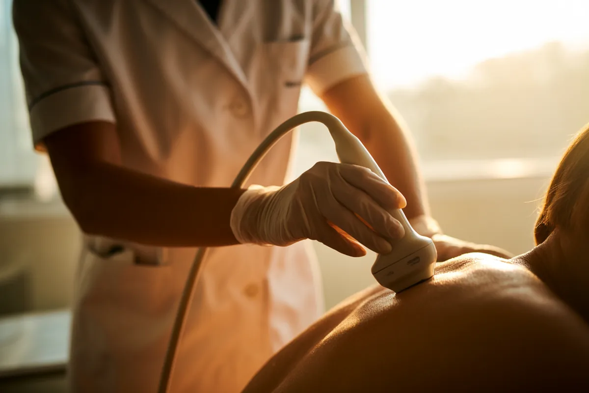
The case for an ultrasound machine in a pain practice is narrower, and more defensible, than the pitch around it. Guidance makes needle placement measurably more accurate, and at some sites safer and less painful to receive. What it has not been shown to do is leave patients feeling better than a landmark injection of the same drug. Buy the machine for accuracy and safety, and teach and document it that way.
Below: the accuracy evidence joint by joint, where the outcome evidence stops, the diagnostic questions that still belong to MRI, the learning curve, and the part most articles skip — who administers which credential, and the documentation and bundling rules. Empire argues the standard-of-care case separately in ultrasound-guided pain injection training and the standard of care; for the wider field, start from the interventional pain procedures reference.
The accuracy gain is real, large, and unevenly distributed
Accuracy is where the evidence is strongest, and it varies by target: the gain is largest where palpation is worst and vanishes where palpation is already good.
| Target | Ultrasound-guided | Landmark-guided |
|---|---|---|
| Hip joint | 100% (95% CI 98–100%) | 72% (56–85%), p<0.0001 |
| Knee joint | 95.8% | 77.8%, OR 6.4 (2.9–14) |
| Biceps tendon sheath | 86.7% | 26.7%, p<0.05 |
| Glenohumeral joint | 92.5% | 72.5%, p=0.025 |
| Subacromial space | 65% | 70% — no advantage |
Two rows carry most of the teaching. The subacromial space — the most commonly injected shoulder target — shows no accuracy advantage. And the knee’s landmark accuracy is approach-dependent: a superolateral approach in extension pools around 91%, an anterolateral approach nearer 67%, so “guidance improves knee accuracy” is really a statement about which blind approach you were using. Small joints of the hand and foot gain most. EULAR puts the principle in a line: accuracy “depends on the joint, route of entry, and health professional expertise.”
Where the outcome evidence stops
The 2021 Cochrane review of guided versus unguided glucocorticoid injection for shoulder pain pooled 19 trials and 1,035 participants. Pain was 0.5 points better with ultrasound on a 0–10 scale (95% CI 0.2 to 0.8, moderate certainty) — below any accepted minimal clinically important difference — the function estimate’s confidence interval crossed zero, and adverse events did not differ. Verbatim: the review “does not support use of image guidance for injections in the shoulder,” and any added cost “appears unjustified.” Non-Cochrane meta-analyses of the same trials disagree, but only because they report those magnitudes as standardized mean differences and call them effectiveness; the dispute is about importance thresholds, not the numbers.
The cleanest single trial is the one to teach from. In 184 patients with inflammatory arthritis, with contrast adjudicating placement, a third of injections guided by clinical examination were inaccurate, and ultrasound-guided injections by a trainee rheumatologist beat examination-guided injections by more senior rheumatologists — 83% versus 66%, P=0.010 — with no significant difference in clinical outcome. Accurate injections, however achieved, did produce greater six-week function improvement, and clinicians using ultrasound could judge their own accuracy while those using palpation could not. That is the honest argument for training: guidance compresses the experience gap and gives real-time feedback.
Guideline bodies split the difference. EULAR says imaging guidance “may be used to improve accuracy” while noting that clinical outcomes are similar. The 2026 multisociety corticosteroid guideline says guidance “increases the accuracy of injections and reduces procedural pain” — not patient-reported outcome — and its companion says imaging improves the safety of stellate blocks, lower-extremity peripheral nerve blocks and some trigger point sites. The ACR’s board-approved 2026 osteoarthritis update summary goes furthest, strongly recommending ultrasound guidance for hip intra-articular glucocorticoid injection on low-certainty evidence; treat it as a board-approved summary with no journal citation yet.
Safety carries the second real signal: across eight trials and 500 wrists, carpal tunnel corticosteroid injection had a lower adverse-event rate with guidance (RR 0.32, 0.21 to 0.49). Two caveats travel with this literature: Cochrane flagged performance and detection bias in most shoulder trials, and several of the largest pro-guidance effects come from trials whose ultrasound arm also used a different syringe and technique, so they do not isolate ultrasound. Guidance also rescues no indication, and a probe lowers no infection risk: aseptic technique still governs.
Diagnostic accuracy: the full- versus partial-thickness split
For rotator cuff tears that split is the whole story. Pooled across 30 studies and 2,402 shoulders, ultrasound gives full-thickness sensitivity 0.91 (0.86–0.94) and specificity 0.93. Across 23 studies and 2,109 shoulders, partial-thickness sensitivity is only 0.68 (0.54–0.83), with specificity 0.94. Never teach a single blended accuracy figure for cuff tears: it conflates the easy case with the hard one. A Cochrane diagnostic review agrees from a surgical reference standard — ultrasound and MRI are statistically indistinguishable for full-thickness tears, and both may be poorly sensitive for partial-thickness tears — and its populations were surgical candidates, so the figures do not transfer unchanged to an unselected clinic.
Structure also does not equal symptoms. In asymptomatic shoulders one series found any cuff tear in 23%, rising from 13% in the fifties to 51% over eighty, while a separate series of asymptomatic people aged 50–79 found full-thickness tears in 7.6% — so quote whichever figure you use with its definition attached, because the unqualified higher number misleads.
ACR Appropriateness ratings map where MRI still wins. Ultrasound rates “usually appropriate” for rotator cuff disorder and subacromial-subdeltoid bursitis (median 8) and for extra-articular hip tendinitis or bursitis (7), and “usually not appropriate” for hip labral tear (2), hip cartilage (1), shoulder labral pathology (2) and knee tendon, meniscus or ligament abnormality (3, against MRI at 9). One safety-critical limit belongs in every curriculum: ultrasound answers is there fluid, and where do I put the needle; it does not answer is this fluid infected. Synovial fluid culture remains the diagnostic test, and in paediatric septic hip the false-negative rate ran about 5%.
Learning curve, and why operator dependence is task-dependent
There is no validated scan count for general musculoskeletal ultrasound competence. A systematic review of 43 studies found only one meeting a high-quality methodological cut-off, with most relying on subjective comfort level. AMSSM says it plainly: “performing a specific number of ultrasound procedures does not necessarily determine competence.”
What exists is task-specific, and the spread is the lesson. Needle visualization plateaued after about five trials on a gelatine phantom but took about 28 supervised trials on cadaveric tissue for the same skill, where only 6 of 15 novices reached proficiency — a six-fold difference driven purely by model fidelity, which is the honest argument against a weekend producing competence. For diagnosis, accuracy for supraspinatus full-thickness tears plateaued between 50 and 75 scans, and the authors recommended about 100 before clinical application. And for ultrasound landmarking before lumbar puncture, 19 attempts were insufficient: four of five fellows never reached 80% success.
Society and program numbers are eligibility thresholds, not competence evidence. EULAR strongly recommends 100 or more prior scans before an intermediate course and 300 or more before an advanced course, and issues no certificate. ACGME’s 2026 physical medicine and rehabilitation requirements set 40 procedures using ultrasound guidance, 10 performed and up to 30 simulated, and for diagnostic musculoskeletal ultrasound only 10 total, none required to be performed — with the guardrail that minimum case numbers “must not be interpreted as equivalent to the achievement of competence.” ACGME’s 2026 rheumatology requirements contain no such requirement at all.
Operator dependence is real, but the blanket version of the claim is refuted and it must be framed as task-dependent. Across 200 shoulders, an MSK-experienced radiologist and a general radiologist with no musculoskeletal ultrasound experience agreed on full-thickness tears 98% of the time (kappa 0.95) and on partial-thickness tears 90% (kappa 0.79) — yet partial-thickness accuracy was poor for both readers. Task difficulty, not operator grade, was the dominant limit. Volume still matters on harder tasks, where high-volume operators outperform occasional ones.
Measurement reliability sets the ceiling: carpal tunnel diagnosis rests on median nerve cross-sectional area thresholds, while that measurement’s inter-rater reliability at the tunnel inlet is poor and published cutoffs vary across reviews — so do not track nerve area across different operators. The training sequence follows: diagnostic competence before guided needling, phantom to cadaveric tissue to supervised patients, and a validated performance assessment rather than a logbook total.
Credentialing: who administers what
The physician and advanced-practice credential is RMSK, Registered in Musculoskeletal, administered by APCA, the Alliance for Physician Certification & Advancement, alongside ARDMS under Inteleos. AIUM does not administer it: AIUM publishes practice parameters and training guidelines and accredits practices in three musculoskeletal specialties. There is no AIUM practice parameter specific to musculoskeletal interventional guidance, so do not cite one, and the freely downloadable AIUM MSK parameter is the 2017 revision, not the current document.
RMSK eligibility is specific and widely misquoted: 150 musculoskeletal ultrasound studies performed or authorized within the preceding 36 months, in clinical diagnostic settings, of which no more than 5% (8 cases) may be therapeutic. The rule that no case may be therapeutic belongs to the separate sonographer credential, RMSKS, administered by ARDMS, which requires two examinations; RMSK does not require the sonography principles examination. Eligible licences include NPs and PAs, applicants keep an auditable case log, and CME is recommended rather than required — 30 MSK-specific credits are suggested as preparation. Maintenance is shifting: CME periods ending on or before December 31, 2026 require 30 credits in musculoskeletal ultrasound, while periods ending on or after December 31, 2027 require 25 credits that need not be specialty specific.
AIUM’s training guidelines ask for 150 studies and 30 AMA PRA Category 1 Credits™ within 36 months for MSK diagnostic, interventional or peripheral nerve practice, at least 50 supervised guided procedures under a separate guideline, and 30 CME with 200 cases per 36 months for maintenance. Read all of these as eligibility requirements: “150 studies is what AIUM and APCA require” is accurate, while claiming research shows 150 studies produces competence is not. One time-sensitive item: the ACR’s RhMSUS certification is currently paused while the ACR evaluates the program’s future and potential transition options, verified on the ACR’s own page on September 17, 2026. AIUM’s guidelines still list it, so do not send a rheumatologist down that route right now.
Medicare does not require a credential to bill. In the policies reviewed — one jurisdiction’s extremity ultrasound coverage determination and billing article, the national coverage determination on diagnostic ultrasound, and the 2026 correct coding manual — the only operator requirement is performance by “qualified and knowledgeable physicians and/or technicians (sonographers) under the general supervision of a physician,” with no credential named. Scope that carefully: coverage determinations are jurisdiction-specific, and privileging, state scope and accreditation are separate levers. In an AIUM-accredited practice, sonographers must be certified even where physicians need not be.
Privileges govern what you may actually do. Authorization comes from your state practice act and your facility’s privileging decision, which weighs training, experience and demonstrated competence. Accredited CME is a legitimate input into that file and nothing more: it does not grant privileges, expand legal scope, or confer subspecialty board eligibility in pain medicine, which at the ABPN and ABPMR requires completing 12 months of ACGME-accredited fellowship training — see CME-accredited pain training versus a pain medicine fellowship. PAs and NPs work through an extra layer — practice act, delegation or collaboration agreement, privileges, payer rules: see whether PAs can perform pain management injections and advanced ultrasound training for physician assistants. Scope is state- and facility-specific; nothing here substitutes for your practice act, supervision agreement or privileging criteria.
Documentation: the report is federal, the image is not
The written report is on the firmest ground. The Medicare Claims Processing Manual states that “the interpretation of a diagnostic procedure includes a written report,” and its professional-component section, citing 42 CFR 415.120(a), requires an interpretation and written report for inclusion in the patient’s medical record — and it sets a quality floor, since a notation reading “fx-tibia” would not suffice.
The permanently recorded image is weaker than it is usually taught: the claims-processing chapter normally cited for it carries no ultrasound image-retention requirement, and neither does the 2026 correct coding manual. It lives in local coverage policy as a “should” — a permanent record of the ultrasound and its interpretation should be kept in the patient’s record, while the “must” is that documentation be available to Medicare on request — and in society parameters, where the multisociety MSK parameter says “there should be a permanent record of the ultrasound examination and its interpretation.” No federal or contractor language ties image retention to the guidance code specifically. The concrete, checkable standard comes from AIUM practice accreditation instead.
AIUM accreditation image checklist for a guided procedure
- All images show the date and are labeled with the injection site and side.
- A labeled image before injection.
- Labeled images during injection: in-plane, “needle shaft and tip must be seen and labeled”; out-of-plane, the tip labeled for clarity.
- The image of the needle is consistent with the description in the report.
- A labeled image after injection that “must demonstrate injectate in the correct location.”
Bundling: the rule that decides most denials
Two statements from the 2026 Medicare correct coding manual carry the lesson. If a code descriptor or instruction indicates that a procedure includes radiologic guidance, a provider “shall not separately report” a guidance code; and CPT codes 20600–20611 are “a family of codes describing arthrocentesis… with or without ultrasound guidance.” So report the ultrasound-guided arthrocentesis code for a joint and you do not also report a separate guidance code for that joint. The same policy appears in every surgery chapter, so it is program-wide.
- One unit per encounter. For ultrasound guidance for needle placement, CPT 76942, CMS allows “one unit of service… at a single patient encounter regardless of the number of needle placements performed.”
- Guidance absorbs its own imaging. Guidance procedures “include all radiological services necessary to complete the procedure.”
- A diagnostic study alongside guidance needs a different region, reportable separately only “if the 2 procedures are performed in different anatomic regions” — and services may not be split across dates to dodge edits.
- One unit per joint, even when surrounding bursae are also treated.
All of this is dated to 2026 material, and the clock runs faster than annually: coding edits update quarterly and local coverage revisions run on their own schedule. Confirm descriptors in a licensed current-year CPT Professional codebook and coverage in your contractor’s current policy before billing. For how procedural mix affects a practice, read pain management procedures and clinic economics against your own payer mix.
Building the skill deliberately
Clinicians who get value from a machine pick a few targets they already inject often, learn to scan those regions to a high standard, add guidance to those procedures only, and document to the accreditation standard from the first case. A credentialing file builds itself that way, one logged and reported study at a time. If the injection foundation needs work first, Empire’s Joint, Extremity and Non-Spinal Injection Training covers the non-spinal targets and is accredited for 6.75 AMA PRA Category 1 Credits™ for the complete in-person hybrid program; the blog explains the knee, shoulder and hip injection curriculum and the evidence for PRP indications and outcomes. For blocks, see the companion guide to nerve blocks in outpatient pain practice.
Train with the probe in your hand
Empire’s Advanced Musculoskeletal Ultrasound Guided Injections course is hands-on ultrasound-guided injection training for clinicians who want scanning and needling repetitions rather than another lecture; curriculum and CME accreditation details are on the course page. Clinicians who already have that foundation progress with Advanced Ultrasound for Pain Management Level II.
See the Advanced MSK Ultrasound course Claim Your Prize!