







There is one sentence that tells you a zygomaticus botox complication has occurred before you have examined anything: "I only got my crow's feet treated, but my smile has changed." Dr. Chris Croley treats that sentence as a diagnostic finding in its own right, and his instruction on hearing it is uncompromising: "Question your injection technique. Review your understanding of the anatomy in that area, and what lines you were actually trying to treat."
This resource is about that last clause — what lines you were actually trying to treat. It is a causal analysis of one specific complication pathway, not an injection site guide. Deliberately, no injection points or coordinates appear below; the mapping question is covered separately in the injection site reference. What follows is why a technically reasonable crow's feet treatment ends up weakening a smile elevator.
The two muscle groups involved
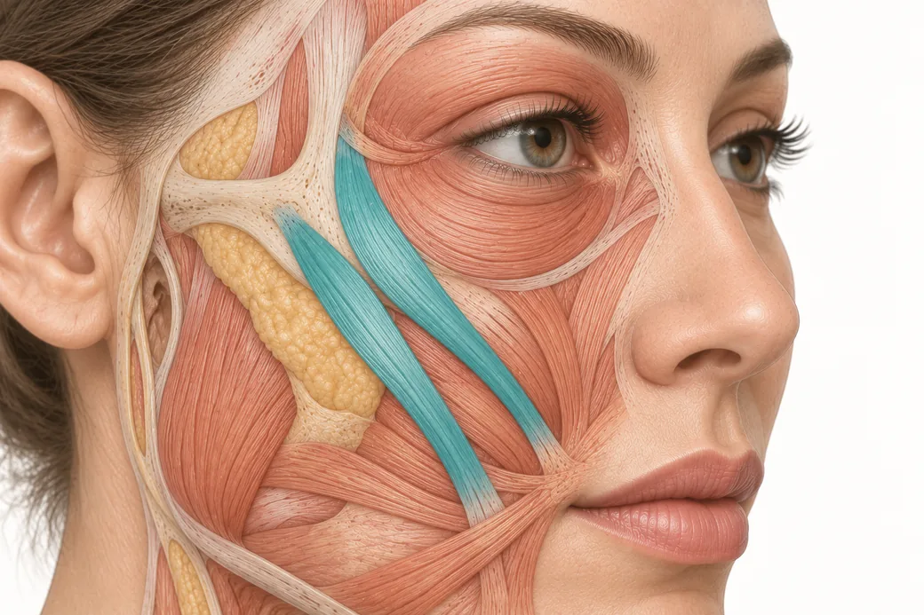
Crow's feet are produced by the lateral fibres of the orbicularis oculi, a superficial sphincter encircling the orbit. This is the intended target, and treating it is entirely standard.
Sitting inferior and deep to those lateral fibres are the zygomatic elevators. Both arise from the zygomatic bone and run obliquely down and medially toward the mouth. Zygomaticus major inserts at the corner of the mouth and elevates and draws the commissure laterally — it is the principal muscle of smiling. Zygomaticus minor runs slightly more medially and inserts into the upper lip, elevating it.
Dr. Croley's summary of why this matters is short: "Those muscles are actually elevators, and they will elevate our smile on one side. So if we stop that muscle movement, this upper lip will not move."
Three features of this arrangement create the trap:
- They are inferior to the target, not lateral to it. The gradient of risk runs downward, in exactly the direction an injector drifts when following a line onto the cheek.
- They are deeper than the target. The orbicularis oculi is superficial. The zygomatici are not. This is a z-axis problem as much as an x-y one.
- There is no barrier. Nothing anatomical stops a deposit intended for the superficial lateral orbicularis oculi from reaching the plane below if it is placed with enough depth, volume or force.
The causal pathway, step by step
This complication is almost never a single dramatic error. It is a sequence of small, individually defensible decisions.
Step one: a correct treatment. The injector treats the lateral orbicularis oculi for crow's feet. Nothing wrong so far.
Step two: inferior creep. Dr. Croley describes exactly where it starts: "As we start moving down into this area and treating kind of the lower portion of the crow's feet, that sometimes we'll start seeing these lateral cheek lines, and we start chasing those lines."
The patient has lines that continue below the orbital rim onto the upper cheek. They look like the same lines. The injector extends the treatment inferiorly to include them.
Step three: going deep. "And especially if we go deep in that area." Lines that persist despite superficial treatment invite a deeper injection, because the injector reasons that the responsible muscle must be deeper. That reasoning is often correct — and the deeper muscle is the elevator.
Step four: a thin patient. "If the patient is really thin, we can impact zygomaticus major or minor." Thin tissue means less distance between planes and less volume to absorb a deposit. The same injection in a patient with more subcutaneous tissue may cause nothing at all, which is precisely why this complication is intermittent rather than universal, and why it takes injectors by surprise.
The outcome: reduced elevation of the upper lip or the oral commissure on the treated side, visible only on smiling, in a patient who was told she was having her eyes treated.
The insight most injectors miss
Notice what step three actually means. If a lateral cheek line is produced by the zygomatic elevators, then treating that line with toxin is not an accident — it is a deliberate weakening of an elevator, performed by an injector who has not realised that is what they are doing.
That reframes the problem usefully. The complication is not primarily a needle-control failure. It is a target-identification failure. The injector set out to treat a line without establishing which muscle produces it, and the muscle that produces it is one you do not want to weaken.
This is the difference between treating a line and treating a muscle. A line is a surface finding — the visible consequence of repeated folding of skin over a contracting muscle, modified by skin quality, volume and photodamage. Some lines in the lateral cheek are produced by muscles you should not touch, and some are not dynamic problems at all, which is where the distinction between dynamic and static findings does real clinical work rather than sitting in a glossary.
Chasing a line to its end is a habit. It is also, in this region specifically, the mechanism of the complication.
Confirming the diagnosis
A patient reporting a changed smile after periocular treatment needs a structured examination, not a reassurance.
Examine with full animation, not at rest. This is a dynamic finding and it may be entirely invisible in a still face. Ask for a full, genuine smile, and repeat it — a posed half-smile will not recruit the elevators fully.
Compare specific structures side to side. Height of the oral commissure at full smile. Elevation of the upper lip. Symmetry of incisor show. The vector and depth of the nasolabial fold, which flattens on the affected side when elevation is reduced.
Localise which elevator. Reduced elevation of the upper lip with relatively preserved commissure position points toward zygomaticus minor. Loss of commissure elevation and lateral draw points toward zygomaticus major. More medial upper lip and nostril involvement suggests the levator group rather than the zygomatici. The distinction matters because it tells you how far medially and how deep your deposit actually travelled.
Check completeness. Partial weakness is a diffusion signature; the muscle received a fraction of a dose. Complete absence of elevation suggests more of the dose arrived than you think.
Consider what else it could be. Not every facial asymmetry appearing after an injection was caused by the injection. New unilateral facial weakness — particularly if it involves the forehead, eye closure and the lower face together, rather than isolated smile elevation — is a different clinical problem and warrants proper neurological assessment rather than being filed as a toxin complication. This is uncommon, and the coincidence of timing is exactly what makes it easy to miss.
Prevention: five boundary rules
Define your inferior boundary before you inject, and hold it. The decision about how far down to treat should be made during planning, when you are thinking about anatomy, not mid-treatment while looking at a line.
Stay superficial laterally. The target is a superficial muscle. There is no clinical reason to go deep in the lateral canthal region, and depth is the single variable that converts this from a safe treatment into a smile complication.
Identify the muscle, then treat it. Before treating any line, name the muscle producing it. If the answer is a zygomatic elevator, toxin is contraindicated for that line in almost every patient who values their smile.
Scale to tissue thickness. A thin patient gets a smaller dose, a smaller volume and a more conservative boundary. Dr. Croley names thin patients specifically as the at-risk group.
Inject slowly and at low volume. Pressure moves drug through planes before diffusion has a say. Force and volume are as relevant here as unit count.
If it has already happened
The prognosis is usually favourable, for a reason that is worth understanding rather than just hoping for. Because this is a diffusion event, the elevator received a fraction of a dose rather than a full one, and the more units reach a muscle the longer it stays weak. The complication therefore tends to resolve ahead of the crow's feet treatment that caused it.
What you cannot do is restore the elevation. Neurotoxin subtracts movement and has no mechanism for adding it back. The only route to symmetry is weakening the corresponding elevator on the opposite side — and Dr. Croley is candid about how that lands: in the correction case he teaches from, the technically successful result was that "even with animation, she has a relatively flat appearance," and "many patients do not want those smile elevators treated."
So the realistic options are a symmetric but flatter smile, or waiting. For most patients without a fixed event on the calendar, waiting is the better trade. Either way, the corrective dose should sit below your usual dose for that muscle, or you will simply move the asymmetry to the other side for longer.
What changes at the chairside
Stop treating the lateral cheek as an extension of the crow's feet zone. It is a different anatomical region with different contents and a different risk profile.
Write your own inferior boundary into your treatment plan, and if a patient has lines below it, have the conversation about why they are not a toxin target rather than quietly extending the treatment.
And when a smile changes after periocular work, run Dr. Croley's instruction rather than reassuring the patient it will pass: question the technique, review the anatomy, and establish which lines you were actually treating. That is how the same complication stops happening to the next thin patient.
This approach reflects Dr. Chris Croley's clinical practice as taught in Empire Medical Training's hands-on curriculum. Technique is learned under supervision; this article is educational and is not a substitute for training.
Plane and depth in the lateral canthal and upper cheek region are difficult to learn from illustrations, because illustrations flatten the dimension that causes the problem. Empire teaches these relationships on tissue in Special Anatomical Cadaver Aesthetics Training, and the periocular region in depth in Master Eye & Nose Injection Training. A general orientation to the surrounding regions is available in the facial mapping reference.
Related guides in this cluster
Part of Facial Muscle Anatomy for Neurotoxin.
 Clinical GuideMost Neurotoxin Complications Are Diffusion, Not Misplacement — Rethinking Root CauseMost neurotoxin complications come from diffusion into an adjacent muscle, not from injecting the wrong site. Here is how to tell which
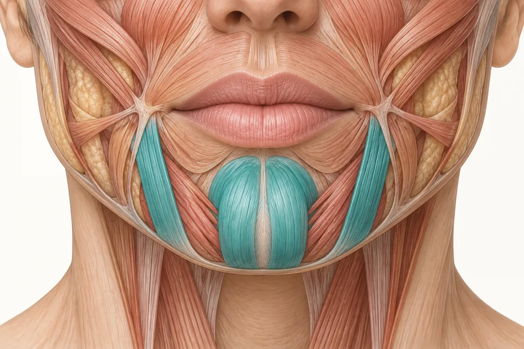 Clinical GuideDepressor Anguli Oris, DLI and Mentalis — Three Overlapping Muscles, Millimetres ApartDepressor anguli oris anatomy is a depth problem, not a map problem. The DAO, DLI and mentalis overlap in layers, and confusing them al
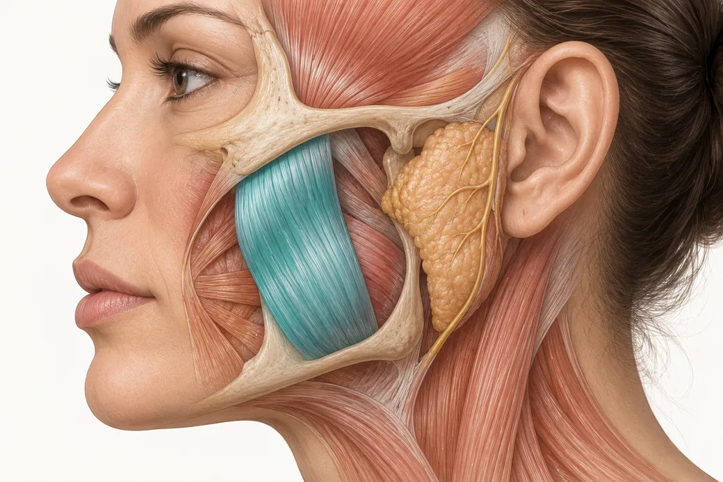 Clinical GuideTwo Clocks, One Muscle: The Masseter Botox Results TimelineThe masseter botox results timeline runs on two clocks — symptom relief in 1–2 weeks, visible facial slimming over months. How to couns
Train with Empire
This guide is clinical education. The technique behind it is taught hands-on, on live patients, with faculty beside you.
Explore Botox Training & Certification →Disclaimer
This article reflects the clinical opinions and experience of Dr. Chris Croley, Chief Medical Officer, Empire Medical Training, an independent faculty member contributing to Empire Medical Training's curriculum. The views expressed are the author's own and do not necessarily represent those of Empire Medical Training.
It is professional education, not medical advice, and is no substitute for hands-on training or independent clinical judgment. Licensed clinicians remain responsible for their own patient selection, technique and outcomes, for verifying current product labelling, and for practising within their scope and applicable law. Empire Medical Training accepts no liability for reliance on this content.
 Claim Your Prize!