








When a patient returns with an unintended effect, almost every injector's first thought is the same: I put the needle in the wrong place. That reflex is understandable, and it is usually wrong. Neurotoxin diffusion complications — dose that leaves the target muscle and reaches a neighbour — account for the large majority of what we call injector error, and the distinction is not semantic. It determines what you actually change about your practice afterwards.
Dr. Chris Croley states the position without hedging: "Usually when we have a complication it's not because I directly injected the wrong muscle. Most of us inject the correct muscles, but often they're very close to other muscles that we may not want to inject. And so occasionally some of the dose that we give leaks over, diffuses over, affects that muscle that we didn't want to treat."
If that is true — and the anatomy says it is — then the standard response to a complication is aimed at the wrong target. Redrawing your injection map does nothing for a problem caused by how the dose behaved after it left the needle.
Why facial anatomy makes diffusion the default failure mode
Facial mimetic muscles are unlike almost every other muscle group a clinician has studied. They are thin. Many are only a few millimetres deep. They insert into skin rather than bone. They overlap, interdigitate, and change plane over short distances. And they are separated from one another not by fascial barriers but, in many places, by nothing at all.
That architecture creates three distinct adjacency geometries, and each produces a different class of complication.
Side-by-side in the same plane. Two functionally opposed muscles sitting beside each other with no meaningful separation. The lateral orbicularis oculi and the upper fibres of the zygomatic elevators are the classic pair. A deposit intended for one sits within diffusion range of the other, and the two do opposite things to the face.
Stacked in depth. Two muscles occupying the same surface coordinates at different depths. Dr. Croley describes exactly this arrangement in the lower face: the depressor anguli oris "is superficial, and right under that is going to be our depressor labii inferioris." Here your map can be perfect and your needle still ends up in the wrong muscle, because the error is in the z-axis, not the x-y plane.
Continuous sheets. Muscles that are not discrete objects at all but regions of a continuous sheet — the frontalis, the orbicularis oculi, the orbicularis oris. There is no edge for the toxin to stop at. Effect grades outward from the deposit, which is why frontalis complications present as gradients of brow position rather than as on-off events.
Once you see the face this way, a complication stops looking like a mistake and starts looking like a predictable consequence of putting a diffusible drug into a crowded, layered, boundary-poor space.
The four things that actually determine spread
If diffusion is the mechanism, then the variables that govern diffusion are the ones worth controlling. Dr. Croley points at three of them directly — needle position measured in millimetres, injection force, and depth — and the fourth, dose, runs through his entire teaching.
Dose. The amount of toxin at the deposit sets how much is available to travel. It also sets how long any spillover lasts, because dose equals duration. A large deposit does not just spread further; the effect it creates next door persists longer.
Volume and pressure. "By treating them, moving our syringe just millimetres, injecting with a little bit too much force, we can drastically alter the smile," Dr. Croley teaches. Force matters because a bolus delivered under pressure is physically pushed through tissue planes before any pharmacological diffusion occurs. The injection technique itself displaces the drug. Reconstitution volume interacts with the same problem from the other direction — a larger volume carrying the same units occupies more tissue on arrival. Dilution strategy is its own subject and is addressed separately in Empire's reconstitution material; the point here is that volume and pressure are anatomical variables, not just pharmacy ones.
Depth. Depth is the most underweighted variable in facial neurotoxin work, and it is the one that converts a correct site into a wrong muscle. Going deep in the lateral cheek to chase a line reaches a different muscle than staying superficial at the same coordinates.
Tissue thickness. "If the patient is really thin, we can impact zygomaticus major or minor," Dr. Croley notes about lateral cheek injections. A thin patient has less soft tissue between planes and less volume to absorb a deposit. The same injection in two patients is not the same injection.
Note what is absent from that list: the location of your injection point on a face chart. That is the reframe. Three of the four drivers of a diffusion complication are things you do at the moment of injection, not things you decided while planning the treatment.
Distinguishing diffusion from genuine misplacement
The two look similar in the mirror and require different responses. Work through these before concluding anything about your technique.
| Signal | Points to diffusion | Points to misplacement |
|---|---|---|
| Distance from your injection point to the affected muscle | Adjacent — same region, within a centimetre or two, or directly deep to the target | Remote from anything you injected |
| Completeness of the unwanted effect | Partial — the muscle still works, just less | Complete, comparable to your intended effect |
| Intended effect | Present and appropriate | Weak or absent, because the dose went elsewhere |
| Duration | Resolves ahead of the intended treatment | Runs the full expected duration |
| Reproducibility across patients | Occasional, patient-dependent, worse in thin patients | Consistent — it happens every time you treat that area |
That last row is the most diagnostically useful one over a career. A diffusion problem is intermittent. A mapping problem is reproducible. If the same unwanted effect appears in most patients you treat in a given region, your understanding of that region's anatomy is the issue and you should go back to it — ideally on a cadaver, where plane and depth are visible rather than inferred. If it appears in one patient in fifteen, and that patient was thin, or you were rushing, or you used a larger volume that day, you have a diffusion problem and the fix lives in dose, depth, volume and pressure.
Three worked examples
Crow's feet reaching the zygomatic elevators. The patient reports that her smile changed after what she describes as a crow's feet treatment. Dr. Croley's account of the mechanism is specific: as the injector moves inferiorly to chase lateral cheek lines, and especially if the needle goes deep, the deposit reaches zygomaticus major or minor. "Those muscles are actually elevators and they will elevate our smile on one side. So if we stop that muscle movement, this upper lip will not move." The injection site was arguably defensible. The depth and the inferior creep were not.
Depressor anguli oris reaching depressor labii inferioris. You intended to weaken the DAO to lift the oral commissure. The DLI sits directly beneath it. Dr. Croley describes the outcome bluntly: "We go from trying to create an upturned smile to having the patient look like they had a stroke." This is the single most disfiguring lower-face toxin complication, and it is a depth error in a region where the correct depth is measured in millimetres.
Frontalis gradients. Because the frontalis is a continuous sheet, a deposit produces a field of effect rather than a discrete one. Asymmetric brow position after forehead treatment is usually a gradient problem — unequal effective dose across the sheet — rather than a missed muscle. This is why frontalis asymmetry is the complication clinicians see most often, and why it is a dosing question more than a placement one.
A fourth example is worth naming even though it sits outside this transcript: eyelid ptosis following glabellar treatment, a long-recognised diffusion event in which toxin reaches the levator palpebrae superioris. It is the textbook case of the principle and is well documented in the product labelling for the on-label glabellar indication.
The post-complication debrief
When a complication occurs, run five questions in order. The order matters, because the first four are about the mechanism and only the fifth is about your map.
- Which muscle produced the unwanted effect? Name it precisely. "The smile is off" is not a diagnosis. Zygomaticus minor is.
- Where does that muscle sit relative to my deposit — beside it, beneath it, or continuous with it? This identifies which adjacency geometry you are dealing with, and therefore which variable to change.
- Was the effect partial or complete? Partial means diffusion. Complete means the dose arrived there.
- What was different about this patient or this injection? Thin tissue, larger reconstitution volume, a faster or more forceful injection, a deeper needle angle, a bigger dose than usual.
- Only now: was my injection point where I intended, and was my intention correct?
Most clinicians start at question five, find that their injection point matched their chart, conclude that they did nothing wrong, and change nothing. The complication then recurs. Starting at question one usually produces an actionable answer — go shallower in the lower face, reduce volume in thin patients, stop chasing lines below the orbital rim.
What this changes on Monday morning
The practical output of this reframe is a short list of habits, not a new map.
- Treat depth as a deliberate decision, not a default. In the lower face and the lateral cheek, decide your plane before the needle enters and be able to say why.
- Scale dose and volume to tissue. A thin patient gets less room for error in every direction. Adjust accordingly.
- Inject slowly. Pressure displaces drug before pharmacology gets a vote.
- Stop treating lines as targets. A line is a surface finding. The muscle beneath it is the target, and chasing a line past the muscle that causes it is how injectors walk into the zygomatic elevators.
- Distinguish a distribution question from a dose question. Whether a region needs more points or a different total is a genuinely different decision from where to put the needle, and the injection site reference and facial mapping guide address the mapping half of that question.
It is also worth being clear about which findings neurotoxin can address at all. A complication caused by diffusion into an elevator affects dynamic movement, and dynamic problems and static ones are different objects — a distinction covered in the dynamic versus static wrinkles discussion.
This approach reflects Dr. Chris Croley's clinical practice as taught in Empire Medical Training's hands-on curriculum. Technique is learned under supervision; this article is educational and is not a substitute for training.
Depth, plane and adjacency are difficult to learn from diagrams, because diagrams flatten exactly the dimension that causes the problem. Empire teaches these relationships on tissue in the Special Anatomical Cadaver Aesthetics Training, and the dosing and technique side is covered in Cosmetic Neurotoxins Training.
Related guides in this cluster
Part of Facial Muscle Anatomy for Neurotoxin.
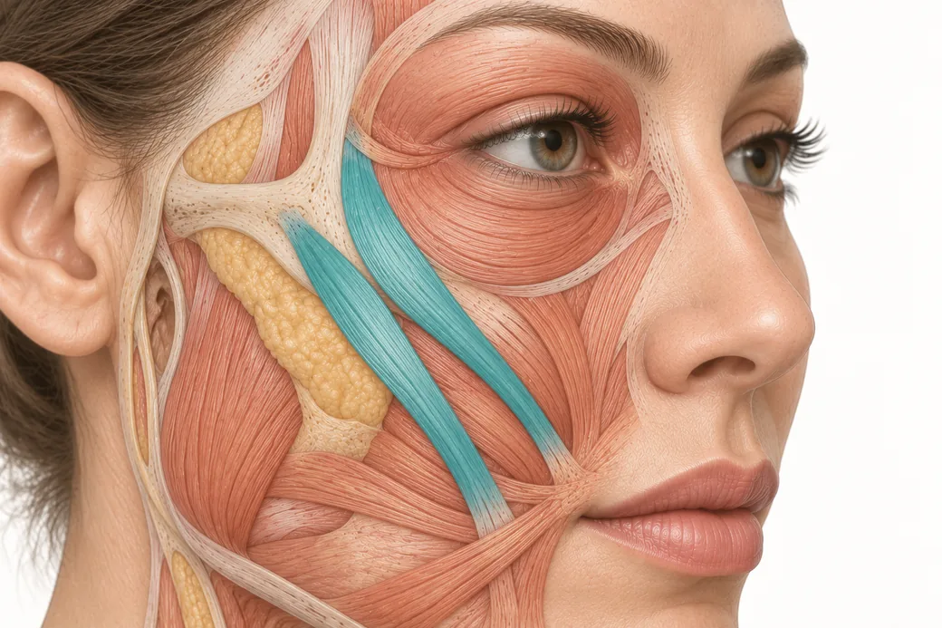 Clinical GuideThe Crow's Feet Trap — How Chasing Lateral Cheek Lines Reaches the ZygomaticusA zygomaticus botox complication after crow's feet treatment has one usual cause — chasing lateral cheek lines too low and too deep. Th
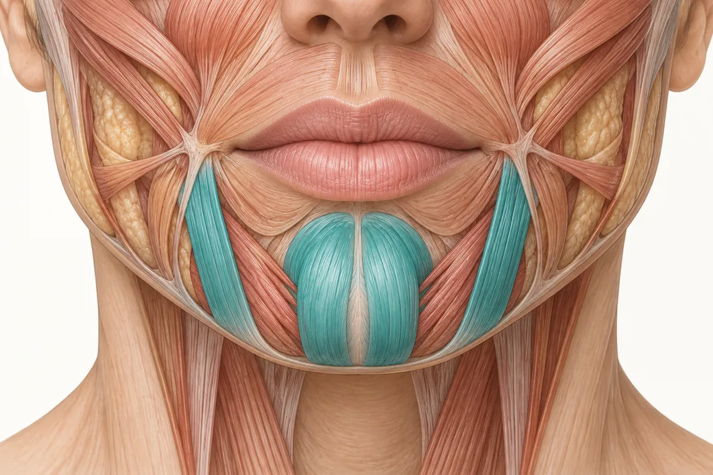 Clinical GuideDepressor Anguli Oris, DLI and Mentalis — Three Overlapping Muscles, Millimetres ApartDepressor anguli oris anatomy is a depth problem, not a map problem. The DAO, DLI and mentalis overlap in layers, and confusing them al
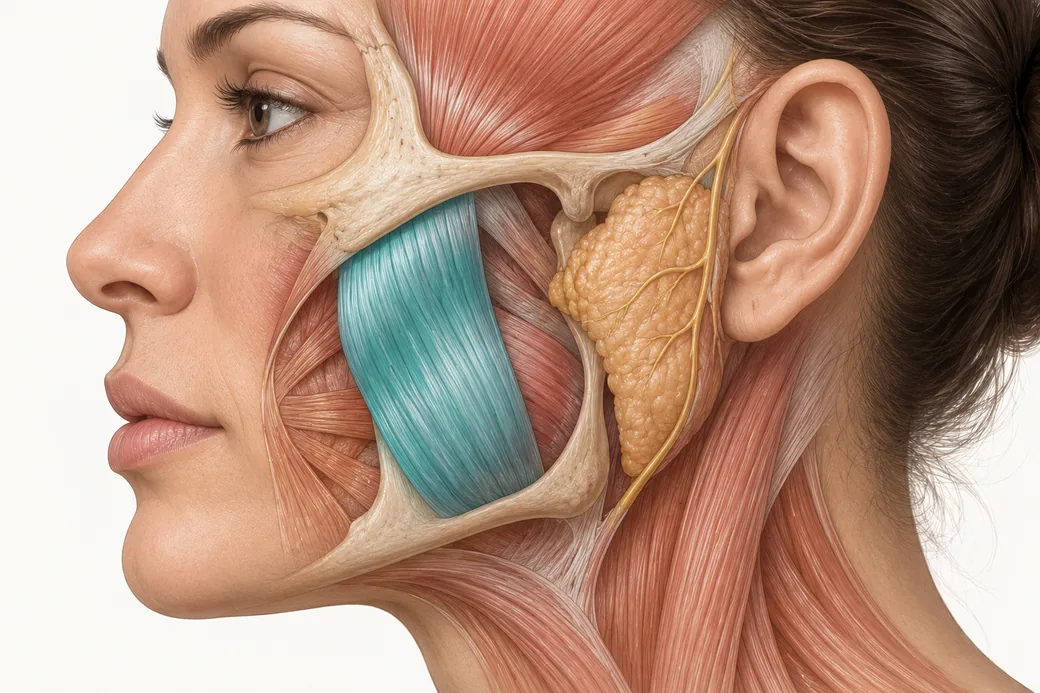 Clinical GuideTwo Clocks, One Muscle: The Masseter Botox Results TimelineThe masseter botox results timeline runs on two clocks — symptom relief in 1–2 weeks, visible facial slimming over months. How to couns
Train with Empire
This guide is clinical education. The technique behind it is taught hands-on, on live patients, with faculty beside you.
Explore Botox Training & Certification →Disclaimer
This article reflects the clinical opinions and experience of Dr. Chris Croley, Chief Medical Officer, Empire Medical Training, an independent faculty member contributing to Empire Medical Training's curriculum. The views expressed are the author's own and do not necessarily represent those of Empire Medical Training.
It is professional education, not medical advice, and is no substitute for hands-on training or independent clinical judgment. Licensed clinicians remain responsible for their own patient selection, technique and outcomes, for verifying current product labelling, and for practising within their scope and applicable law. Empire Medical Training accepts no liability for reliance on this content.
 Claim Your Prize!