







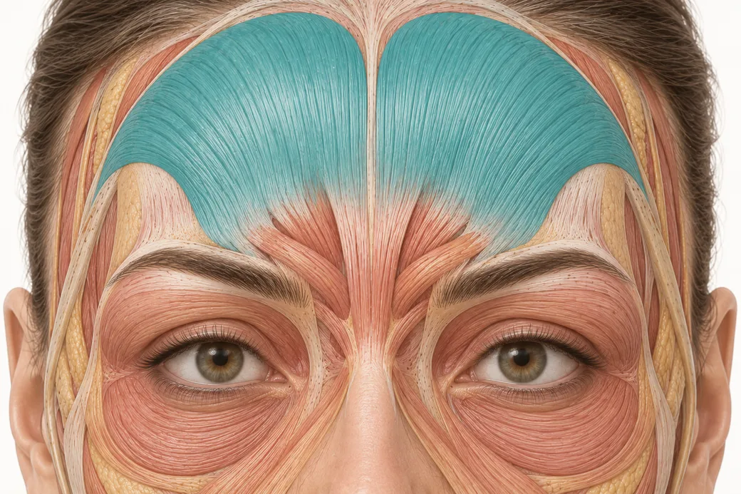
A spock brow after botox is almost never a placement accident and almost never diffusion. It is compensation — the predictable behaviour of muscle fibres that were left working while their neighbours were switched off. Melissa Pulcini-Buttine, PA names the mechanism in a single sentence: "If you only treat medially in the frontalis, you're leaving all of this lateral portion on, it's really unaffected. So you may relax the medial part, but then the lateral part is going to overcompensate. And that's when you get spocking or quizzical eyebrows."
Understanding it as compensation rather than error changes what you do about it, because the two have opposite fixes. An error is corrected by being more careful. Compensation is corrected by treating more muscle, not less.
What the patient is actually showing you
The presentation is specific and worth describing precisely, because it is frequently miscalled. The lateral end of the brow sits sharply higher than the medial end, producing an unnaturally peaked arch: the Mephistopheles effect in the surgical literature, spocking or the quizzical brow in the clinic. The peak is usually at or just lateral to the brow apex, and it is exaggerated on animation because animation is when the recruited fibres do their work.
Two features distinguish it from the things it gets confused with. First, it is usually bilateral and roughly symmetric — compensation follows the treatment pattern, and the treatment pattern was applied to both sides. Asymmetric sharp elevation is a different problem. Second, the medial brow is often not merely un-elevated but actively lower than baseline, and this is the finding that identifies the mechanism. The published description of the Mephistopheles effect attributes it to placement of medial and especially lateral injection points high above the corrugator supercilii, catching the frontalis and producing medial brow depression with compensatory lateral brow elevation (Aesthetic Surgery Journal Open Forum 2025;7:ojaf032).
In other words: two things happened, not one. Something went down and something else came up in response.
The mechanism, in order
Step one: a territory is relaxed. Either the medial frontalis is treated directly, or glabellar injections placed high catch the medial frontalis fibres. The medial brow loses elevator input and settles.
Step two: the lateral frontalis is not relaxed. It may have been deliberately spared — the commonest reason, usually as a precaution against brow ptosis — or simply not reached by the pattern used.
Step three: the lateral fibres recruit. This is the part that surprises people. Muscle does not politely maintain its previous level of activity when a neighbouring segment stops working. Facial movement is goal-directed: the patient is still trying to raise the brows, still trying to widen the eyes on animation, and the fibres capable of doing it take on more of the work. The published consensus describes it as uneven muscle immobilisation causing excessive recruitment of unrelaxed fibres, and lists among its consequences both accentuation of previously unnoticed wrinkles and aesthetically unpleasing lateral brow elevation.
Step four: the contrast makes it worse. A lateral brow elevating against a medial brow that has descended produces a larger angular difference than either change alone. The patient perceives one dramatic change; you caused two moderate ones in opposite directions.
Why cautious injectors produce it more often
The uncomfortable part of this pattern is that it is generated by exactly the instinct that prevents a worse complication.
An injector who is properly afraid of brow ptosis will stay high and stay medial. Staying high is correct. Staying medial is the error, and it is an error that feels like prudence. It comes from treating "how much muscle do I dare relax?" as the safety question, when the safety question is actually "which fibres will still be working when I am finished, and what will they do?"
Melissa's framing of the frontalis makes the trap visible: the whole muscle is one elevator with one job, and treating a fraction of it does not produce a fraction of the effect. It produces a different effect, with a different shape. Her instruction — "respect the movement of this muscle and inject accordingly" — is an instruction about the whole muscle, not about restraint in one part of it.
There is a second contributor, which is that the lateral extent of the frontalis is genuinely hard to know. It has no bony landmarks. It arises from the galea aponeurotica rather than bone, and its lateral border sometimes inserts into the superior temporal septum. Frontalis line patterns come in four described types, and in around 15 percent of people the lines form two lateral columns with no central component at all — a forehead in which the lateral fibres are the dominant ones and a medially-weighted pattern is close to the worst possible choice.
Prevention: three decisions, made before you inject
Decide the lateral border by watching, not guessing. Have the patient raise the brows maximally and watch where forehead movement actually stops. The extent, severity and location of dynamic rhytids let you estimate the shape and boundaries of that patient's frontalis; it is an inference, but it is a patient-specific one, and it is far better than a fixed distance from the midline. If the lines run out laterally at one point in one patient and two centimetres further in the next, your treated territory should differ by the same amount.
Limit by dose and height, never by territory. This is the single most useful reframing in the piece. If a patient's brow cannot tolerate full frontalis relaxation — a compensated brow, an older patient with poor tissue support, a heavy lid — the correct restraint is fewer units spread across the whole width, and placement kept high. The wrong restraint is a full-strength medial treatment with the lateral third untouched. The first produces a softer, evenly relaxed forehead. The second produces the spock.
Check what the glabellar pattern is doing to the medial frontalis. Since medial brow depression is half of the mechanism, glabellar points placed too high are a contributing cause. The depth logic for the corrugator's medial head and lateral tail is covered in a companion resource in this cluster and is directly relevant here: a lateral corrugator injection placed too deeply or too high reaches frontalis fibres it was not meant to reach.
Management once it has happened
The corrective principle follows from the mechanism: you treat the fibres that are doing the over-elevating.
Published management is to treat the apex of muscle elevation on the frontalis with low-dose injections, identified by watching where the peak movement occurs rather than by measuring from the brow. Consideration can also be given to the relevant brow depressors — procerus, depressor supercilii, orbicularis oculi and its palpebral part — where the aim is to bring the lateral brow down rather than to raise the medial one.
Three constraints on that:
- Dose low. You are correcting a shape, and the muscle you are treating is already partly the muscle you want working. The failure mode of over-correction is a flat, heavy brow, which is worse than a peaked one.
- Wait the full interval. Toxin continues to set in for up to two weeks, and a brow assessed at day five is a moving target. General timing of corrective intervention is covered elsewhere in Empire's neurotoxin material; the point here is simply that a spock brow at day six may be a different shape at day fourteen.
- You cannot raise the medial brow. If the medial descent is substantial, the honest conversation is that toxin is a subtractive drug — the only available move is bringing the high side down, not lifting the low side up.
The consultation consequence
Patients returning with a quizzical brow frequently arrive convinced their injector "put it in the wrong place." The more accurate and more reassuring explanation is that the treated area was too small rather than misplaced, and that the correction is additional treatment rather than reversal. That conversation is easier if the possibility was raised beforehand, and raising it costs one sentence at the consultation: that relaxing part of a muscle can make the untreated part work harder, that it is adjustable, and that the adjustment happens at the two-week review rather than immediately.
For clinicians building the regional picture, Empire's botox face chart and botox injection sites overviews are the reference layer for where these areas sit, and botox dosages for the forehead and frontalis area covers the dosing side of the restraint decision described above.
Recognising compensation patterns as a class — rather than memorising each one — is what anatomy-led training is for. Empire's cosmetic neurotoxins training and anatomical based aesthetics training both work through muscle-by-muscle functional relationships of this kind.
The clinical reasoning above reflects Melissa Pulcini-Buttine's practice as taught in Empire Medical Training's hands-on curriculum. Technique is learned under supervision; this article is educational and is not a substitute for training.
Melissa Pulcini-Buttine, PA is a physician assistant of two decades and has been a professor of anatomy and physiology for approximately fourteen years. She is a faculty member at Empire Medical Training and the founder of an aesthetics practice in Greenwich, Connecticut.
Related guides in this cluster
Part of Facial Muscle Anatomy for Neurotoxin.
 Clinical GuideMost Neurotoxin Complications Are Diffusion, Not Misplacement — Rethinking Root CauseMost neurotoxin complications come from diffusion into an adjacent muscle, not from injecting the wrong site. Here is how to tell which
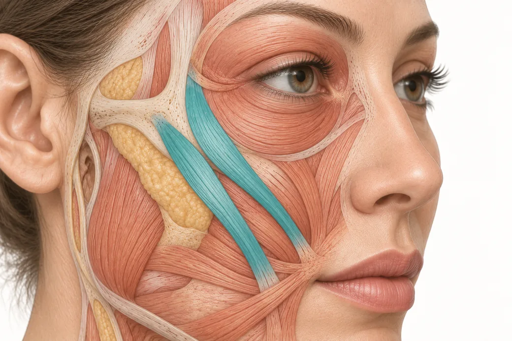 Clinical GuideThe Crow's Feet Trap — How Chasing Lateral Cheek Lines Reaches the ZygomaticusA zygomaticus botox complication after crow's feet treatment has one usual cause — chasing lateral cheek lines too low and too deep. Th
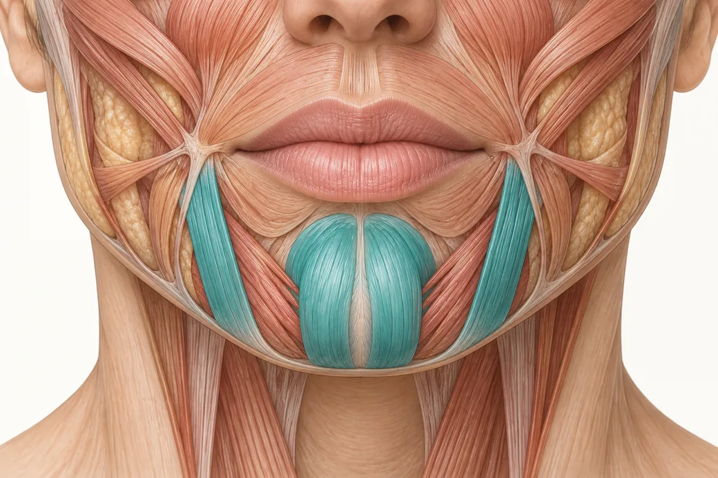 Clinical GuideDepressor Anguli Oris, DLI and Mentalis — Three Overlapping Muscles, Millimetres ApartDepressor anguli oris anatomy is a depth problem, not a map problem. The DAO, DLI and mentalis overlap in layers, and confusing them al
Train with Empire
This guide is clinical education. The technique behind it is taught hands-on, on live patients, with faculty beside you.
Explore Botox Training & Certification →Disclaimer
This article reflects the clinical opinions and experience of Melissa Pulcini-Buttine, PA, an independent faculty member contributing to Empire Medical Training's curriculum. The views expressed are the author's own and do not necessarily represent those of Empire Medical Training.
It is professional education, not medical advice, and is no substitute for hands-on training or independent clinical judgment. Licensed clinicians remain responsible for their own patient selection, technique and outcomes, for verifying current product labelling, and for practising within their scope and applicable law. Empire Medical Training accepts no liability for reliance on this content.
 Claim Your Prize!