







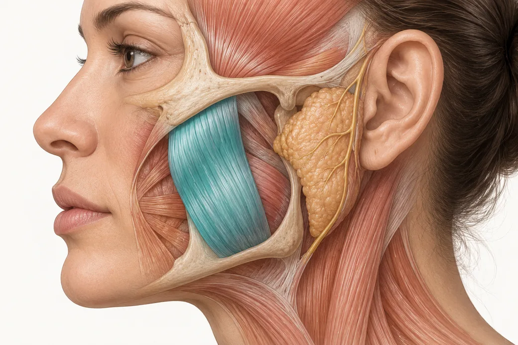
The masseter injection safe zone is not a decorative marking exercise. It is the difference between a treatment that narrows a jawline and one that produces an asymmetric smile, a dry mouth or a bulge the patient can see every time they bite down. When we map this out, what we are really doing is thinking about the muscles around the area we are treating — because this is neurotoxin, and with neurotoxin you have to treat the right muscles.
Surface anatomy is the tool that makes that possible. This piece is the map: what the masseter actually is in three dimensions, how to draw its boundaries on a live face, what sits immediately outside those boundaries, and why depth matters as much as position.
The masseter is not one muscle belly
Most injectors carry a mental image of the masseter as a single rectangular slab on the side of the jaw. It is layered, and the layers do different things.
The muscle is conventionally described in three layers, fused anteriorly and diverging posteriorly:
- The superficial layer arises from a thick aponeurosis on the temporal process of the zygomatic bone and the anterior two-thirds of the inferior border of the zygomatic arch. Its fibres run inferoposteriorly, passing over the deep portion, and insert onto the angle of the mandible at the masseteric tuberosity and the lower lateral surface of the ramus.
- The intermediate layer arises from the middle third of the zygomatic arch and blends with the superficial layer anteriorly and at its mandibular insertion.
- The deep layer arises from the deep surface of the zygomatic arch, runs inferiorly, and inserts higher on the lateral ramus than the superficial portion.
Functionally, all three elevate the mandible and approximate the teeth. The intermediate and deep fibres retract the mandible; the superficial fibres protrude it. Innervation throughout is the masseteric nerve, a branch of the mandibular division of the trigeminal nerve, CN V3, which reaches the muscle from its deep surface through the mandibular notch (Anatomy, Head and Neck, Masseter Muscle, StatPearls, NCBI Bookshelf). More recent cadaveric work has argued that the conventional three-part description is itself incomplete, describing a distinct coronoid part of the human masseter (Mezey and colleagues, Annals of Anatomy, 2021).
Two structural details matter enormously for injection:
Obliquity. The fibres run obliquely, not vertically. A needle entering perpendicular to the skin crosses fibre planes rather than following them, which is one reason depth and distribution behave less predictably here than in a thin muscle of facial expression.
Internal tendon. Within the superficial head there is a substantial internal tendinous structure — described in the complication literature as the deep inferior tendon — that divides the superficial head into superficial and deep bellies. It is a physical barrier to spread, it varies in thickness and morphology between patients, and it is directly implicated in uneven toxin distribution. In a retrospective case-control analysis, muscles that developed paradoxical bulging had significantly thicker deep inferior tendons than controls, 0.85 mm versus 0.60 mm, and compartment-type or transverse-type tendon morphology carried higher risk than longitudinal type (Sun et al., Journal of Cosmetic Dermatology, 2026).
This is the anatomical reason the masseter cannot be treated as a bag of muscle into which toxin is deposited and allowed to find its own way.
These figures reflect Dr. Croley's clinical practice as taught in Empire Medical Training's hands-on curriculum. Technique is learned under supervision; this article is educational and is not a substitute for training.
Drawing the boundaries on a live face
The marking sequence I teach is short and it is done with the patient clenching, because you cannot find the borders of a relaxed masseter reliably.
First, the superior boundary. Draw a line from the tragus to the oral commissure. This is the single most important mark on the face for this treatment, and it functions as a ceiling. Published safe-zone descriptions use exactly this landmark: the line connecting the oral commissure and the tragus is the upper boundary of the generally recommended injection region, and injections are placed below it to reduce the risk of diffusion into adjacent facial muscles (Yoshida, Toxins, 2026). Some authors use the inferior border of the earlobe rather than the tragus as the posterior anchor; the intent is identical, which is to capture the bulk of the masseter below the line while keeping the needle away from the zygomaticus complex above it.
Second, the anterior and posterior borders. With the patient clenching, palpate and mark the anterior and posterior edges of the contracting muscle. These are patient-specific; do not assume them from a diagram.
Third, the inferior boundary. The line of the mandible. Mark the inferior border, and mark the antegonial notch — the concavity in the inferior mandibular border just anterior to the angle — as your anterior-inferior reference point.
Those four marks define a quadrilateral. The published safe zone is described the same way: bounded by the tragus-to-commissure line above, the jaw line below, and the anterior and posterior borders of the masseter on each side.
With those marks you have delineated the muscle. Now find where its bulk actually is. On the great majority of patients, the bulk of the masseter sits low — in the inferior portion of that quadrilateral, over the angle and the lower ramus. That is where the treatment goes. The bulk of the treatment belongs where the bulk of the muscle mass lies.
This is also broadly consistent with what is known about motor endplate distribution. The motor endplate zone of the masseter is concentrated in a band-like region within the central portion of the muscle belly, often corresponding to the lower-central region of the clinically palpable muscle — which is to say, the same region palpation already identifies (Yoshida, Toxins, 2026).
What sits above the line, and why it ends careers
The tragus-to-commissure line is a ceiling because the anatomy above and anterior to it is expressive.
Zygomaticus major and zygomaticus minor. Both arise from the zygomatic bone and descend anteroinferiorly toward the mouth — zygomaticus major to the modiolus at the oral commissure, zygomaticus minor to the upper lip. They are the principal elevators of the corner of the mouth. Weaken them and the patient's smile drops on that side. They lie above the line, anterior and superior to the masseter's upper portion, which is precisely why going too high in the face with a masseter injection is a technique error rather than a stylistic choice.
Risorius. A thin, variable muscle running horizontally to the modiolus, superficial and anterior to the masseter's anterior border. It is the muscle most often blamed when a masseter patient develops an asymmetric smile, and its horizontal course means it sits close to the anterior portion of the tragus-to-commissure line.
Depressor anguli oris and the lateral orbicularis oris. Both are within diffusion range of an over-anterior injection.
The published risk statement is explicit: excessively anterior injection increases the risk of diffusion into the risorius, zygomaticus major and minor, depressor anguli oris and the lateral portion of the orbicularis oris, potentially causing smile asymmetry or other unwanted facial changes (Yoshida, Toxins, 2026). In the largest published complication series — 680 patients across 2,036 masseter treatment sessions — smile limitation occurred after 0.15% of sessions (Peng and Peng, Journal of Cosmetic Dermatology, 2018). Rare, and entirely a placement problem.
I do not go up to that line at all. The line is where you stop caring about it, not where you aim.
The other two boundaries have their own hazards
Anteriorly: vessels and expression. The anterior border of the masseter is where the facial artery and facial vein cross the inferior border of the mandible to enter the face. Published guidance names the extreme anterior border as a region to avoid specifically because of facial vessel injury, in addition to the expressive-muscle diffusion risk. Injectors who are careful about vascular anatomy in filler work — the same discipline behind our material on whether botox can cause blindness and vascular safety generally — should carry that carefulness across to the anterior masseter border.
Posteriorly and superiorly: the parotid. The parotid gland overlies the posterior portion of the masseter, and Stensen's duct crosses the muscle superficially before turning medially at its anterior border. Posterior-superior placement risks diffusion into the gland, and xerostomia appears among the reported injection-site complications of masseter neurotoxin. Avoid the posterior-superior region near the parotid.
The practical margin. Complication-prevention guidance from the large-series literature is to keep injections inside the safe zone and ideally in three to four separate locations at least 1 cm from any border. That single sentence is a complete technique rule, and it accounts for most of what goes wrong.
Depth: the error that is unique to this muscle
This is a deep muscle. A very common error is to treat it superficially, and it is the error that produces visible sequelae rather than merely a weak result.
Practical depth estimates in the literature place superficial injections at approximately 5–10 mm and deeper injections at approximately 15–20 mm, with the explicit caveat that these are practical estimates rather than fixed rules and must be individualised to subcutaneous tissue thickness and muscle volume (Yoshida, Toxins, 2026). The point that matters at the chairside is that depositing the whole dose above the deep inferior tendon, leaving the deep fibres innervated, is a recognised route to paradoxical bulging.
The response in the complication literature is a dual-plane technique: three injection points to the inferior half of the masseter, ensuring toxin is placed in both the deep and the superficial plane of the muscle (Ozsoy and colleagues, Aesthetic Surgery Journal Open Forum, 2024). Needle length matters here in a way it does not in the upper face — a short 4 mm or 6 mm needle chosen for forehead work cannot reach the deep belly of a thick masseter.
The technique this map supports
My own approach follows directly from the marking. I dose about 20 units per masseter, and I divide it across approximately three injection points rather than depositing the whole dose at one site. The bulk of the dose goes low, on the jawline but on the anterior surface of the muscle, with the remainder placed at the midpoint of the muscle. I do not go up to the tragus-to-commissure line. In a male patient with a genuinely prominent masseter — or a female patient with a large one — two additional injection points can be added to cover a bigger muscle.
The reason I prefer division over a single bolus is distribution. Some providers place the whole dose at one point and allow it to diffuse through the muscle. In my experience that is how you get strange movements: the superficial fibres are affected, the deep fibres are not, and when the patient clenches, the muscle bulges.
For context rather than for copying: published masseter dosing clusters at 20–40 units per side, and one review concluded that doses below 20 units per side are inadequate (Ghatge et al., Bioinformation, 2023). Twenty units sits at the lower end of that range. Dose selection is a separate discussion from mapping, and it belongs with the rest of a practitioner's dosing philosophy rather than with surface anatomy.
What this changes at the chairside
- Mark every masseter, every time, with the patient clenching. Tragus to oral commissure. Anterior border. Posterior border. Mandibular line. Four marks, thirty seconds.
- Treat the inferior half. Bulk of the dose where the bulk of the muscle is, at least 1 cm inside every border, and nowhere near the line.
- Check your needle length before you draw up. A masseter is not a glabella, and the upper-face habits that inform most botox injection sites and standard botox face chart mapping do not transfer to a muscle this deep.
Masseter marking, depth and injection are taught hands-on in Empire's Cosmetic Neurotoxins Training, with the regional anatomy covered in Anatomical Based Aesthetics Training and dissected in Special Anatomical Cadaver Aesthetics Training. For the therapeutic indication, see our material on botox for jaw clenching.
About the author
Dr. Chris Croley — Chief Medical Officer, Empire Medical Training. The clinical approach described here is what he teaches in Empire's hands-on neurotoxin curriculum.
Related guides in this cluster
Part of Facial Muscle Anatomy for Neurotoxin.
 Clinical GuideMost Neurotoxin Complications Are Diffusion, Not Misplacement — Rethinking Root CauseMost neurotoxin complications come from diffusion into an adjacent muscle, not from injecting the wrong site. Here is how to tell which
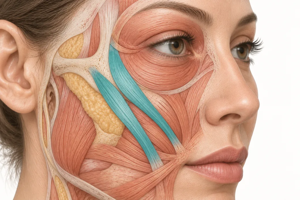 Clinical GuideThe Crow's Feet Trap — How Chasing Lateral Cheek Lines Reaches the ZygomaticusA zygomaticus botox complication after crow's feet treatment has one usual cause — chasing lateral cheek lines too low and too deep. Th
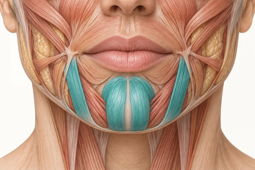 Clinical GuideDepressor Anguli Oris, DLI and Mentalis — Three Overlapping Muscles, Millimetres ApartDepressor anguli oris anatomy is a depth problem, not a map problem. The DAO, DLI and mentalis overlap in layers, and confusing them al
Train with Empire
This guide is clinical education. The technique behind it is taught hands-on, on live patients, with faculty beside you.
Explore Botox Training & Certification →Disclaimer
This article reflects the clinical opinions and experience of Dr. Chris Croley, Chief Medical Officer, Empire Medical Training, an independent faculty member contributing to Empire Medical Training's curriculum. The views expressed are the author's own and do not necessarily represent those of Empire Medical Training.
It is professional education, not medical advice, and is no substitute for hands-on training or independent clinical judgment. Licensed clinicians remain responsible for their own patient selection, technique and outcomes, for verifying current product labelling, and for practising within their scope and applicable law. Empire Medical Training accepts no liability for reliance on this content.
 Claim Your Prize!