








The brow ptosis vs eyelid ptosis distinction is the most consequential differential in neurotoxin practice, and it is routinely collapsed — by patients, understandably, and by injectors, less so. The patient calls and says "my eye is drooping." That sentence contains no diagnostic information at all. It is compatible with two entirely different complications, arising from two different muscles, with two different nerve supplies, two different mechanisms of causation, two different management pathways and two different recovery timelines.
Getting the differential right matters for three reasons: it tells you what you actually did wrong, it determines whether there is anything useful you can offer the patient, and it changes what you do differently at the next injection. Getting it wrong means you adjust a technique that was not the problem.
"We need to know our muscles, we need to know the depths, and we need to make sure the angle of our needle is pointing the correct way," says Melissa Pulcini-Buttine, PA, who has taught anatomy and physiology for roughly fourteen years. The reason all three matter is that the two ptoses are produced by failures in three different variables — and only an anatomical model tells you which one failed.
The two muscles
Frontalis. The frontalis is the only elevator of the eyebrow. That is the entire clinical story and it deserves to be stated starkly, because injectors treat it as one muscle among many when it is functionally a solo act. It has no bony origin; it arises from the galea aponeurotica and inserts into the skin and the fibres of the brow depressors. Its antagonists are the depressor group — procerus, corrugator supercilii, depressor supercilii, and the superior fibres of orbicularis oculi. Innervation is the temporal branch of the facial nerve, CN VII.
Weaken the frontalis and nothing else lifts the brow. The depressors, now unopposed, win. The brow descends. The upper lid skin, which sits on the brow, descends with it — and the patient, looking in the mirror, reports a heavy, hooded, drooping eye. This is brow ptosis, and the eyelid itself is entirely normal.
Levator palpebrae superioris. This is a different muscle in a different compartment doing a different job. It originates from the lesser wing of the sphenoid at the orbital apex, runs forward through the orbit above the superior rectus, and inserts via its aponeurosis onto the tarsal plate of the upper eyelid. Its job is to elevate the eyelid itself. Innervation is the superior division of the oculomotor nerve, CN III — not the facial nerve.
Weaken the levator and the lid margin drops, independent of where the brow is. This is true eyelid ptosis.
Two muscles, two cranial nerves, two compartments. Melissa's point about knowing "what their origin is, what their action is, and what their antagonistic muscles do" is not an academic exercise here. The origin tells you why one muscle is reachable by a subcutaneous forehead injection and the other is not. The antagonist relationship tells you why weakening one elevator produces a descent you did not inject for.
How each one actually happens
Brow ptosis: an injection that was too low, or too much
Brow ptosis is a dosing-and-position failure in the frontalis, and it is almost always the inferior frontalis — the portion close to the brow — that causes it.
The logic is mechanical. The frontalis acts on the brow at its inferior insertion. Weaken the muscle where it does its lifting and you remove the lift. Weaken it high on the forehead and you soften horizontal lines while leaving the lifting portion functional.
This is the anatomical basis of a rule Melissa teaches that every injector eventually learns the hard way: if you want elevation, stay high.
"If we want elevation we're going to stay high above that line of convergence," she teaches. "If you want more elevation in a region, stay higher up, stay more superior. If you want to drop her brow, then you could go more straight across and you would have a more straight appearance in the eyebrows."
She works from the line of convergence, a landmark identified dynamically rather than by measurement — the patient raises the brows and you observe the level at which the direction of frontalis movement reverses. Skin displacement analysis has demonstrated that reversal in every volunteer studied (Cotofana S, Freytag DL, Frank K, et al. Plastic and Reconstructive Surgery. 2020;145(5):1155–1162). Injecting above the line preserves lift; treating too far below it removes the lift and produces descent. "If you're worried about true eyebrow ptosis, that's when you come too low. Now the patient can't elevate, because the job of the frontalis is to elevate — but now you're going to cause depression there."
The practical rule is entirely standard: the inferior frontalis is where brow ptosis is manufactured. Approaching frontalis dosing systematically is covered in Botox dosages for the forehead and frontalis area.
There is a second and less obvious route to brow ptosis: over-treating the frontalis in a patient who was using it compensatorily. Some patients with pre-existing brow descent or dermatochalasis are chronically recruiting frontalis at rest to keep the visual field clear. They do not report this and are often unaware of it. Relax that muscle and you have not caused a new problem — you have unmasked an existing one. This is why static assessment before treatment matters, and why Melissa insists on "really looking at the patient staying still, and then the movement" before marking anything.
True eyelid ptosis: toxin that travelled
True eyelid ptosis is a diffusion failure. You did not inject the levator palpebrae superioris — it sits inside the orbit, well beyond the reach of any aesthetic injection. What happened is that product placed near the superomedial orbital rim migrated into the orbit and reached it.
Melissa identifies the specific anatomical conduit she teaches injectors to respect: "You have a little foramen in here, the supraorbital foramen — a little hole where there's an artery but also a little nerve coming out of there. So we don't want any of our toxin to get into there and potentially come inferiorly and affect the levator palpebrae. Then we would get a true eyelid ptosis."
She is equally direct about the mechanical error that delivers it there, and it is not depth — it is angle. "If I was pointing laterally, I could spread my toxin into the no-go zone and cause a true eyelid ptosis."
That is the part injectors under-appreciate. An injection at the correct site, at the correct depth, with the correct dose, delivered with the needle angled toward the orbit, deposits product on a trajectory heading where you do not want it. The three variables Melissa names — muscle, depth, angle — are independent, and angle is the one nobody photographs.
The other contributing factors are the ordinary ones: excess volume at a single point, over-dilute reconstitution increasing spread, injecting too close to the orbital rim, and massage or manipulation of the area shortly after treatment.
The chairside differential
The patient cannot tell you which one it is. These tests can.
1. Compare the brow to baseline. This is why pre-treatment photography is not a marketing asset but a diagnostic one. If the brow has descended relative to baseline, brow ptosis is present or contributory. If the brow is where it was, it is not.
2. Look at the lid margin, not the skin fold. In true eyelid ptosis the upper lid margin itself sits lower relative to the pupil and the corneal limbus. In brow ptosis the lid margin is in its normal position — what has descended is the brow and the redundant skin draping over it. Train yourself to look at the margin and ignore the fold, because the fold is what the patient is describing.
3. The manual brow-lift test. Gently elevate the patient's brow with your thumb to its pre-treatment position and ask what they see. If the heaviness resolves completely and the eye looks normal, the problem was the brow. If the lid margin still sits low with the brow held up, the levator is involved.
4. Test frontalis function. Ask for a brow raise. Absent or markedly reduced elevation confirms the frontalis is down. Note that this does not exclude a concurrent true ptosis — the two can coexist, and in a heavily treated upper face they sometimes do.
5. Look for compensatory recruitment. In isolated true eyelid ptosis with an intact frontalis, patients often over-recruit the frontalis on the affected side to lift the lid, producing a raised brow and a furrowed forehead unilaterally. That pattern — low lid, high brow — is close to diagnostic, and it is the opposite of the brow ptosis picture.
6. Note the timing. Brow ptosis tends to appear as the rest of the toxin effect appears. True ptosis is more often reported somewhat later in the first week or ten days, as diffusion continues. This is a clinical pattern rather than a reliable rule and should support the diagnosis rather than make it.
Why the distinction changes what you can offer
Brow ptosis has no pharmacological antidote. There is nothing to reverse; the muscle is simply weakened and will recover on the ordinary timeline of the product used. What you can sometimes do is rebalance — selectively reducing the activity of the brow depressors so the residual frontalis function is less opposed. That is an antagonist-based decision and it requires knowing which depressor is doing what, which is precisely why Melissa teaches antagonist relationships rather than injection points. It is also a judgement call in an already-treated face, and doing it badly compounds the original error.
True eyelid ptosis likewise cannot be reversed — the toxin effect at the levator will run its course. But the eyelid has a second, smaller elevator that the toxin has not touched: the superior tarsal muscle (Müller's muscle), which is smooth muscle under sympathetic control rather than somatic control. Topical alpha-adrenergic agonist eye drops stimulate it and produce a small amount of lid elevation — typically enough to be cosmetically and functionally meaningful while the toxin effect resolves. Apraclonidine 0.5% is the agent most commonly used for this purpose. It is a temporising measure, not a cure, it requires an appropriate prescription and ocular assessment, and it does not shorten the duration of the complication.
That asymmetry is the practical payoff of the differential: for one of these complications there is something you can do, and for the other there is not. Misdiagnose and you either offer a patient a treatment that will not help them or fail to offer one that would.
What changes at the next injection
If it was brow ptosis, the correction is positional and dose-related in the frontalis: stay higher, respect the inferior frontalis, and assess for compensatory recruitment before treating. The treatment plan changes, not the safety protocol.
If it was true eyelid ptosis, the correction is about spread — volume per point, reconstitution concentration, distance from the orbital rim, and above all needle angle. Melissa's instruction to watch "where your needle is pointing, and the angle" is the specific remediation, and it is worth practising deliberately rather than assuming.
And in both cases the underlying correction is the same one she returns to throughout: stop thinking in injection points and start thinking in muscles. "Before the needle goes in, visualise the structures underneath." Mapping tools such as a Botox face chart are useful for organising a plan, but the plan has to be built on what the muscle does, what its antagonist does, and what happens to the balance between them when one side of that pair is switched off. The distinction between the movement you are treating and the surface result you are chasing is the subject of dynamic vs static wrinkles.
These techniques and landmarks reflect Melissa Pulcini-Buttine's clinical practice as taught in Empire Medical Training's hands-on curriculum. Technique is learned under supervision; this article is educational and is not a substitute for training.
Clinicians who want to build neurotoxin practice on dissected muscle anatomy will find it in Empire's anatomical based aesthetics training and special anatomical cadaver aesthetics training. Core toxin competence, including complication recognition, is covered in cosmetic neurotoxins training.
Melissa Pulcini-Buttine, PA — physician assistant of two decades; professor of anatomy and physiology for ~14 years; faculty member, Empire Medical Training; founder of an aesthetics practice in Greenwich, Connecticut.
Related guides in this cluster
Part of Facial Muscle Anatomy for Neurotoxin.
 Clinical GuideMost Neurotoxin Complications Are Diffusion, Not Misplacement — Rethinking Root CauseMost neurotoxin complications come from diffusion into an adjacent muscle, not from injecting the wrong site. Here is how to tell which
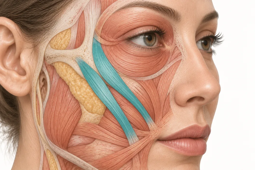 Clinical GuideThe Crow's Feet Trap — How Chasing Lateral Cheek Lines Reaches the ZygomaticusA zygomaticus botox complication after crow's feet treatment has one usual cause — chasing lateral cheek lines too low and too deep. Th
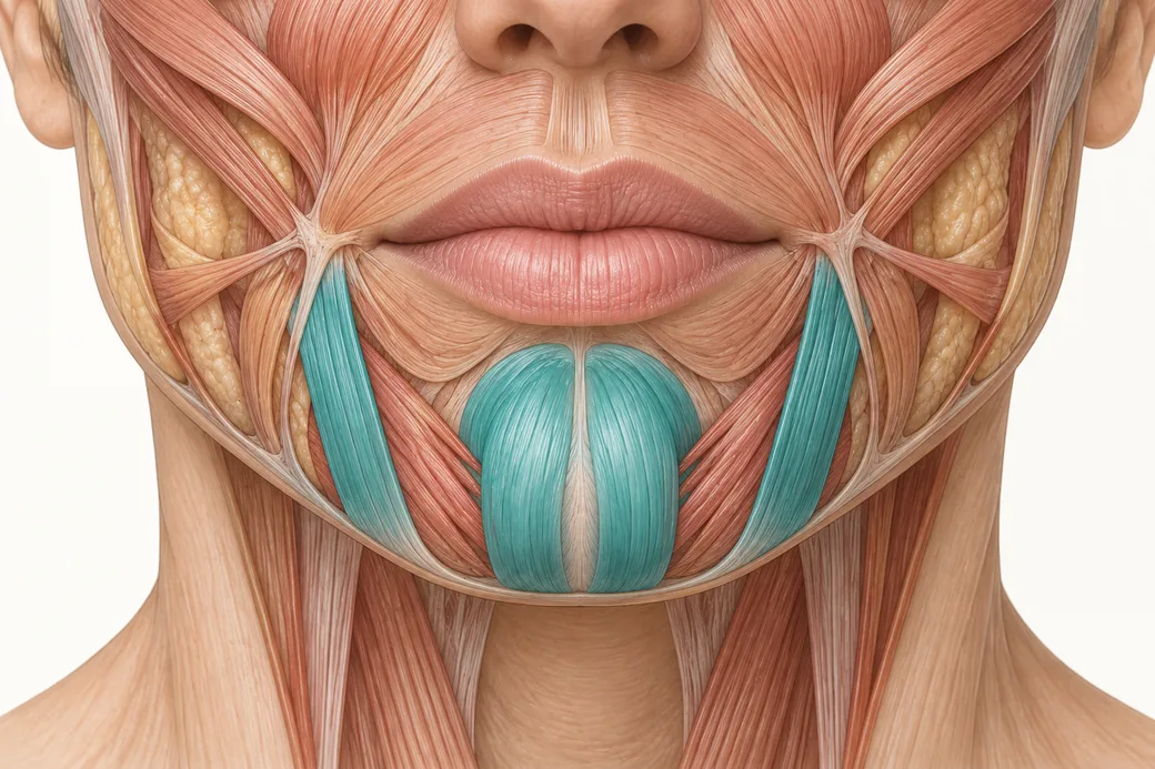 Clinical GuideDepressor Anguli Oris, DLI and Mentalis — Three Overlapping Muscles, Millimetres ApartDepressor anguli oris anatomy is a depth problem, not a map problem. The DAO, DLI and mentalis overlap in layers, and confusing them al
Train with Empire
This guide is clinical education. The technique behind it is taught hands-on, on live patients, with faculty beside you.
Explore Botox Training & Certification →Disclaimer
This article reflects the clinical opinions and experience of Melissa Pulcini-Buttine, PA, an independent faculty member contributing to Empire Medical Training's curriculum. The views expressed are the author's own and do not necessarily represent those of Empire Medical Training.
It is professional education, not medical advice, and is no substitute for hands-on training or independent clinical judgment. Licensed clinicians remain responsible for their own patient selection, technique and outcomes, for verifying current product labelling, and for practising within their scope and applicable law. Empire Medical Training accepts no liability for reliance on this content.
 Claim Your Prize!