








Lower face neurotoxin risk is not a matter of how carefully you inject. It is structural, and it is the reason the same injector with the same steadiness of hand can work comfortably in the glabella and should approach the perioral region as a different discipline. Melissa Pulcini-Buttine, PA states it as a working principle: "Upper face is actually way more forgiving than the lower face."
She is not saying the lower face is dangerous in the vascular sense — that is a separate subject with its own resources. She is saying something more specific and more useful: the amount of error the anatomy will absorb before the patient notices is much smaller below the commissure, and the consequences of exceeding it are qualitatively different. "Lower face toxin is one of my favourites," she says. "Very important anatomy that you need to know in the lower face, because there's a lot of unforgiving spots there. You need to be in the right muscle. You need to be in the right depth."
This resource is about the forgiveness gradient itself: what makes a region tolerant or intolerant of error, why the lower face sits at the intolerant end, and what a rational injector does differently as a result. The layered anatomy of the depressor anguli oris, depressor labii inferioris and mentalis — how those three muscles stack and overlap millimetre by millimetre — is covered in a companion resource in this cluster and is not repeated here.
What "forgiving" actually means
A region forgives error to the extent that a small placement mistake produces a small, correctable, private consequence. It punishes error to the extent that the same mistake produces a large, uncorrectable, public one. Four factors set where a region falls on that scale.
How far apart the muscles are. In the upper face, the functionally opposed muscles occupy distinct territories and, importantly, distinct planes. The glabellar depressors sit deep; the orbicularis oculi is a thin sheet adherent to the skin. Melissa describes it as a muscle so superficial that treatment is effectively intradermal — "like you're doing a bleb." The vertical separation between the structures you want and the structures you do not is measured in millimetres of obvious difference. In the perioral region, the same separation is measured in millimetres of similarity.
Whether the muscles are single-purpose. Upper face muscles do a small number of discrete things. Perioral muscles contribute to composite movements — speech, smiling, eating, drinking, oral competence — that recruit several muscles simultaneously. Weakening one component of a composite movement does not reduce the movement; it distorts it.
Whether an error is visible only to the patient. An upper-face error shows up in a photograph or a mirror. A lower-face error shows up every time the patient speaks. It is visible to their colleagues, their family and their clients, and it is visible in exactly the region observers use to read emotion.
Whether the error can be corrected forward. This is the decisive one. Most upper-face shape errors are correctable by treating more muscle — a peaked brow is managed by treating the apex of elevation, a residual line by treating the fibres that produced it. Almost no lower-face functional error can be corrected by adding toxin to the affected muscle, because the problem is that the muscle is already too weak. The only available manoeuvre is to weaken its opposite number to match, and that means deliberately making a functioning side worse in order to restore symmetry.
That asymmetry between "correct forward" and "balance backward" is the whole of the forgiveness gap.
The specific anatomy that removes the margin
Three structural facts do most of the work, and all three are documented rather than impressionistic.
Everything converges on one point. The modiolus is a thick fibro-muscular band at the oral commissure formed by the convergence of zygomaticus major, depressor anguli oris, levator anguli oris, buccinator, orbicularis oris and risorius (Toxins 2026;18(8):332). Six muscles with different and frequently opposite jobs meet in a structure a few millimetres across. Toxin placed near it does not choose.
The muscles overlap in depth rather than sitting side by side. The depressor anguli oris originates just lateral to the mentum from the oblique line of the mandible, runs upward above and lateral to the depressor labii inferioris, converges into a narrow fasciculus at the oral commissure and inserts into the modiolus. The DAO and DLI partially overlap inferiorly, with the DLI lying beneath the DAO. Platysma fibres reach the lateral border of the DAO and continue to run underneath it. This is a stack, not a map, and the distances are small: ultrasound measurement places the DAO's depth at roughly 4.7 to 5.8 mm at its medial margin and 8.0 to 8.8 mm at its lateral margin, with a thickness declining from about 2.9 mm near the modiolus to about 2.1 mm near the mandibular margin.
The target may not be where you think it is — or there at all. The same ultrasound work found that close to the modiolus the DAO is smaller, and that in nearly one-third of patients no DAO muscle fibres were observed at that location at all. Injecting there risks both a failed treatment and involvement of the other muscles attaching nearby: risorius, zygomaticus major, orbicularis oris, DLI and buccinator.
That last finding reframes the usual advice. The reason to avoid injecting close to the commissure is not only that neighbouring muscles are near. It is that in a substantial minority of patients, the muscle you were aiming at is not present there.
The complication that defines the region
Melissa describes the outcome that makes the lower face different, and she describes it in the terms patients use rather than clinical ones:
"The issue is there's something here called the depressor labii inferioris. So DLI. And if you accidentally hit that muscle, you're going to have an asymmetrical smile. And the patient will almost look like they had a stroke. And it wears off slowly over the next few months, but they're usually not very happy."
Three properties of that complication are worth separating, because each one contributes to the severity.
It is functional, not cosmetic. One side of the lower lip stops depressing. The smile becomes asymmetric in a way that reads as neurological rather than aesthetic, which is why patients and their families reach for the word stroke.
Its duration is not negotiable. It resolves on the toxin's timeline — months, not weeks — and nothing shortens it. There is no hyaluronidase equivalent for neurotoxin.
Its correction requires making something else worse. Melissa is honest about the ceiling: "It's very advanced injectors — we can compensate other muscles to kind of hide it." Compensation means weakening the intact side so the two match. It works, it is a legitimate manoeuvre, and it is not a repair. Meanwhile the corrected side, being treated later and often at a different dose, may outlast or underperform the original, which is how a single asymmetry becomes a sequence of them.
The consensus literature arrives at the same place from a different direction: of the experts surveyed on DAO treatment, 92.8 percent preferred intramuscular injection, typically at a single point lateral to the marionette line, specifically in order to remain within a safe treatment zone and avoid diffusion to the DLI.
The mentalis constraint
The chin deserves separate mention because its risk profile is different again. The mentalis is the only elevator of the lower lip and chin and provides the primary vertical support for the lower lip. Its upper fibres run horizontally and intermingle superiorly with the inferior margin of the orbicularis oris; its lateral fibres descend obliquely and partially intermingle with the DLI.
So the chin presents two distinct hazards on opposite sides of the same muscle. Too deep or too superior and you are into the DLI's territory with the consequences described above. Too much of the mentalis itself and you have weakened the structure that holds the lower lip up, which produces its own functional complaint. Melissa's blunt summary of why depth decides this — "that DLI, which I do not want to hit, is a very deep muscle" — is the operative relationship; the depth values themselves belong to the hands-on curriculum rather than to an article.
The commoner, less dramatic chin problem — residual dimpling at the two-week review — is covered separately, as is the differential between muscular dimpling and volume loss. Empire's existing overview of botox chin dimpling is the patient-facing reference for the region.
Why the upper face tolerates what the lower face does not
It is worth stating the contrast explicitly, because the point is not that the upper face is safe.
Upper-face errors are real and they matter. Brow ptosis is a genuine complication and it also lasts months. The difference is in the category of consequence. A brow that sits too low is a shape problem in a region where shape is the entire purpose of the treatment, and it is at least partially manageable by adjusting the opposing muscles. A lower lip that will not depress on one side is a movement problem in a region whose purpose is movement, and the only management is symmetric weakening.
There is also a difference in the size of the target. The frontalis is a broad sheet several centimetres in every dimension. Being a centimetre off within it changes the distribution of an effect. In the perioral region a centimetre is the difference between two muscles with opposite functions.
This is why the traditional training sequence — upper face first, perioral later, with supervision — is not gatekeeping. It reflects the fact that the lower face has a smaller error budget and a worse failure mode, and that both of those are properties of the anatomy rather than of the injector.
What changes at the chairside
- Treat the perioral region as a different procedure, not a further region. Separate consent, separate consultation time, separate photography. Empire's guidance on what to include in botox consent forms is the right starting point, and asymmetric smile deserves naming explicitly rather than sitting inside a general adverse-event clause.
- Ask what is underneath before choosing depth, every time. Melissa's instruction generalises beyond the chin: "It's really important not just to understand the name of the muscle and the movement, but what's around it, and pick the correct depth to go at."
- Stay away from the commissure. Both because neighbouring muscles converge there and because the muscle you are targeting may be attenuated or absent at that location in a meaningful fraction of patients.
- Treat concentration as a lever, not an afterthought. Melissa is explicit that volume and spread are part of the safety plan in this region alongside placement and units. The specific reconstitution she teaches for the lower face is taught in the hands-on curriculum, where the dilution can be prepared and the spread observed rather than described.
- Start conservatively and use the review visit. A treatment that is slightly weak at two weeks is an easy conversation. A treatment that is slightly too strong in this region is a three-month conversation.
- Photograph in animation, not at rest. A resting perioral photograph will not document the baseline asymmetry the patient already had, and baseline asymmetry is what you will be accused of causing.
The lower face rewards exactly the kind of anatomical knowledge that is difficult to acquire from diagrams, because the relationships that matter are relationships in depth. Empire's anatomical based aesthetics training and special anatomical cadaver aesthetics training address that directly — injecting, then dissecting to see where the product actually went — and the perioral material sits within advanced botulinum toxin and filler training. Melissa's own argument for that format is unadorned: "A lot of times you're going to see, oh wow, I went too deep here and I hit the DLI."
The clinical reasoning above reflects Melissa Pulcini-Buttine's practice as taught in Empire Medical Training's hands-on curriculum. Technique is learned under supervision; this article is educational and is not a substitute for training.
Melissa Pulcini-Buttine, PA is a physician assistant of two decades and has been a professor of anatomy and physiology for approximately fourteen years. She is a faculty member at Empire Medical Training and the founder of an aesthetics practice in Greenwich, Connecticut.
Related guides in this cluster
Part of Facial Muscle Anatomy for Neurotoxin.
 Clinical GuideMost Neurotoxin Complications Are Diffusion, Not Misplacement — Rethinking Root CauseMost neurotoxin complications come from diffusion into an adjacent muscle, not from injecting the wrong site. Here is how to tell which
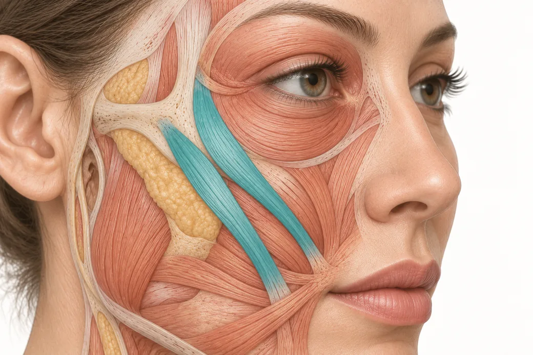 Clinical GuideThe Crow's Feet Trap — How Chasing Lateral Cheek Lines Reaches the ZygomaticusA zygomaticus botox complication after crow's feet treatment has one usual cause — chasing lateral cheek lines too low and too deep. Th
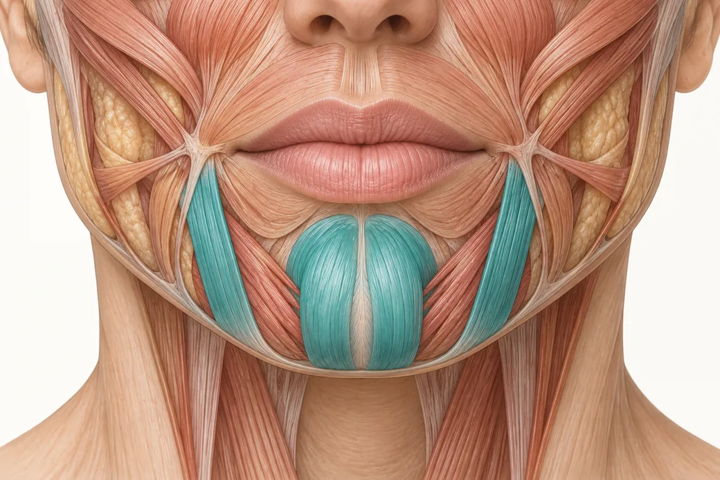 Clinical GuideDepressor Anguli Oris, DLI and Mentalis — Three Overlapping Muscles, Millimetres ApartDepressor anguli oris anatomy is a depth problem, not a map problem. The DAO, DLI and mentalis overlap in layers, and confusing them al
Train with Empire
This guide is clinical education. The technique behind it is taught hands-on, on live patients, with faculty beside you.
Explore Botox Training & Certification →Disclaimer
This article reflects the clinical opinions and experience of Melissa Pulcini-Buttine, PA, an independent faculty member contributing to Empire Medical Training's curriculum. The views expressed are the author's own and do not necessarily represent those of Empire Medical Training.
It is professional education, not medical advice, and is no substitute for hands-on training or independent clinical judgment. Licensed clinicians remain responsible for their own patient selection, technique and outcomes, for verifying current product labelling, and for practising within their scope and applicable law. Empire Medical Training accepts no liability for reliance on this content.
 Claim Your Prize!