







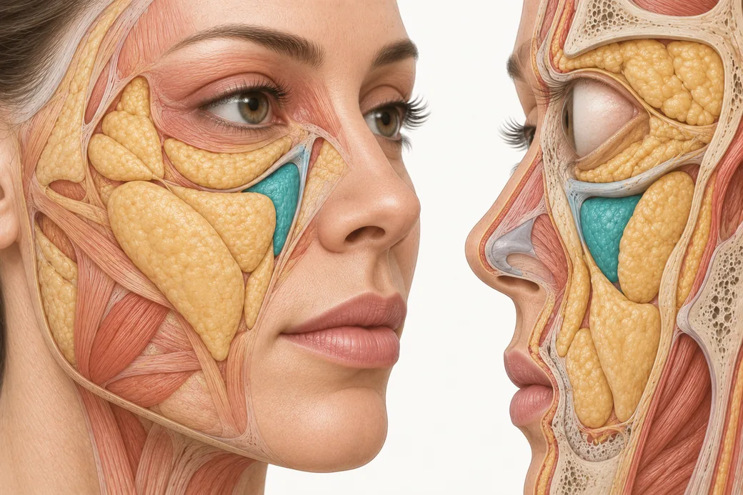
The four-layer model of facial aging is not a teaching diagram. It is a diagnostic sequence — bone, fat, muscle and ligament, skin — and the order matters, because each layer is the foundation for the one above it. Run it deep to superficial and the model does something a list of findings cannot: it tells you which layer the patient's complaint actually lives in, and therefore which modality has any chance of reaching it.
I have taught anatomy and physiology for about fourteen years, and the single most useful thing I can hand an injector is this: aging is layered, so assessment has to be layered too. A patient does not present with a wrinkle. They present with the visible output of four simultaneous processes running at different rates, and the wrinkle is the last and most superficial of them.
This page is the reference for that model. It covers what is documented to change in each layer, what the evidence actually supports, and how to convert the model into an operating procedure at the chairside.
Why the sequence runs deep to superficial
Think of the face as a structure, not a surface. Bone provides projection. Fat provides volume and contour. Muscle and ligament provide suspension and movement. Skin is the covering that drapes over all three, and it shows you everything that has happened underneath it.
If you assess in the opposite direction — surface first — you find the finding that is easiest to see and hardest to fix. The crease is visible. The deficit that created it is not. Assess deep to superficial and you find the driver before you find the complaint, which means you choose your tool once instead of three times.
There is a second reason for the order. The layers are not independent. When bone at the pyriform aperture recedes, the soft tissue that was sitting on it has less platform, so the nasolabial region deepens without any change in the fat or the skin. When a retaining ligament loosens, fat that was held in place descends, and the fold it creates is a position problem rather than a volume problem. Treating the fold as a volume problem in that patient produces a heavier face, not a younger one.
Layer 1 — Bone: the platform that moves
For most of the last century the working assumption was that the adult facial skeleton was stable and that aging was a soft-tissue event. Three-dimensional CT analysis over the past two decades has overturned that. The facial skeleton remodels throughout adult life, and the remodeling is selective: specific regions resorb in a specific and reasonably predictable pattern while adjacent regions stay stable.
The orbit. The bony orbital aperture enlarges with age, and the enlargement is not concentric. Kahn and Shaw, using three-dimensional reconstructions of facial CT scans, documented recession concentrated at the superomedial and inferolateral aspects of the rim, while the central portions of the superior and inferior rims remained comparatively stable (Kahn DM, Shaw RB. Aesthetic Surgery Journal. 2008;28(3):258–264). Pessa and Chen had earlier described the same phenomenon as progressive distortion of the orbital aperture curve on skull specimens (Pessa JE, Chen Y. Plastic and Reconstructive Surgery. 2002;109(2):751–755).
Clinically, that pattern is what produces the aged periorbital signature — a lengthened lid–cheek junction, increased prominence of the medial fat pad, elevation of the medial brow — in a patient whose eyelid skin may be unremarkable.
The midface. Shaw and Kahn measured the glabellar, pyriform and maxillary angles on CT across three age bands and found the glabellar and maxillary angles decreased significantly with age, with the pyriform aperture area increasing (Shaw RB, Kahn DM. Plastic and Reconstructive Surgery. 2007;119(2):675–681). Mendelson and colleagues quantified maxillary retrusion on standardized parasagittal slices through the mid-orbit and found the maxillary angle decreased by roughly 10 degrees between subjects under 30 and subjects over 60 (reviewed in Mendelson B, Wong CH. Aesthetic Plastic Surgery. 2012;36(4):753–760).
The maxilla is more susceptible to age-related loss than the zygoma. That asymmetry between two adjacent bones is the anatomical reason the midcheek deflates medially while the lateral malar region holds its projection longer — and it is why medial midface support so often improves a complaint the patient localized somewhere else entirely.
The mandible. Shaw and colleagues found no significant change in bigonial width or ramus breadth across age groups, while ramus height, mandibular body height and mandibular body length decreased and the mandibular angle increased (Shaw RB, et al. Plastic and Reconstructive Surgery. 2010;125(1):332–342). The obtuse angle and the shortened vertical dimension are what soften a jawline that used to be sharp, independent of any jowl fat.
The mechanism that connects bone to everything above it. As the periosteum retrudes with the bone beneath it, the periosteal attachments of the facial retaining ligaments and mimetic muscles move with it. Those structures do not merely lose a platform; they lose mechanical advantage (Mendelson and Wong, 2012). This is the reason bone is layer one rather than a footnote. A skeletal change of a few millimetres propagates upward into ligament position, muscle vector and eventually surface contour.
An honest note on the evidence. Almost all of this literature is cross-sectional: young subjects compared with old subjects, not the same subject followed over time. Fourgeot and colleagues addressed that gap directly by comparing two facial CT scans in the same individuals a minimum of seven years apart (Aesthetic Surgery Journal. 2021;41(12):NP1907–NP1915). Intra-individual data of that kind remain limited, and the magnitude of skeletal change in any given patient in front of you is not something you can predict from a population mean. Use the pattern to know where to look. Do not use it to quote a number to a patient.
Layer 2 — Fat: compartments, not a blanket
The second layer is where most injectors already think they are working, and where the anatomy is most frequently oversimplified. Subcutaneous facial fat is not a continuous sheet. Rohrich and Pessa demonstrated on thirty hemifacial cadaver dissections that it is partitioned into discrete anatomical compartments with reproducible boundaries — the nasolabial fat as a discrete unit, what had been loosely called "malar fat" separating into medial, middle and lateral temporal-cheek compartments, three forehead compartments, three orbital compartments, and jowl fat as the most inferior (Plastic and Reconstructive Surgery. 2007;119(7):2219–2227).
Those boundaries are real structures. Subsequent histologic work showed the septa are fibrous condensations running from superficial fascia into the dermis, and that some of what has been described as a "retaining ligament" is simply the fusion point of two abutting septal barriers (Rohrich RJ, Pessa JE. Plastic and Reconstructive Surgery. 2008;121(5):1804–1809). Schaverien and colleagues then showed each compartment is associated with an identifiable perforating vessel travelling in those same septal boundaries (Plastic and Reconstructive Surgery. 2009;123(2):695–700) — which is why compartment anatomy is a vascular subject as much as a volumetric one.
The practical consequence: the face does not deflate as a unit. It deflates compartment by compartment, and adjacent compartments can move in opposite directions at the same time.
What the imaging literature actually agrees and disagrees on. Be careful here, because the published data are genuinely in conflict. Cevik Cenkeri and colleagues, using repeat MRI in 70 patients a median of 44.5 months apart, found volume and thickness decreased significantly in all three medial superficial cheek compartments, with width decreasing superiorly and increasing inferiorly — a descent signature (Dermatologic Surgery. 2020;46(12):1600–1605). The overlapping group's larger repeat-CT study of 262 patients found something partly different: total superficial cheek fat volume and the inferior compartment volume increased, while the upper and middle compartment volumes decreased (Sarigul Guduk S, et al. Journal of Cosmetic Dermatology. 2022;21(4):1430–1435).
Both studies agree on the direction that matters clinically — superior compartments lose, inferior compartments gain width and height, and the fat layer as a whole descends. They disagree on whether total volume falls. In the lower face the pattern is clearer still: Guo and colleagues, controlling for BMI, found the inferior superficial jowl and deep jowl compartments thickened with age while the deep labiomandibular layer thinned (Plastic and Reconstructive Surgery. 2024;153(3):539e–548e).
So "volume loss" is an incomplete description of the fat layer. Redistribution is the better word, and at the chairside it means the question is never only how much but where, and in which direction. Our consumer-facing overview of facial volume loss is a reasonable page to send a patient to; it is not the model you should be assessing with.
Superficial versus deep. The compartments are also stacked. Deep compartments sit against bone or deep fascia and behave as structural volume; superficial compartments sit in the subcutaneous layer and move with the skin. Ultrasound work by Schelke and colleagues quantified that difference in living faces: on smiling, the superficial midfacial compartments moved an average of 3.7 mm cranially (Journal of Cosmetic Dermatology. 2021;20(12):3849–3856). A compartment that translates several millimetres every time the patient smiles is not the same target as one that does not, and that distinction belongs in your plan before you choose a product.
Layer 3 — Muscle and ligament: the suspension system
The third layer is the one most likely to be skipped, and it is the layer that explains position rather than volume.
Retaining ligaments. The facial retaining ligaments anchor the superficial musculoaponeurotic system and the overlying skin to the underlying skeleton and deep fascia. Age-related laxity of these ligaments is a recognised contributor to facial sagging, jowling and volume descent, and the anatomy is still being refined — Park and Noel recently used high-resolution sectioned images and three-dimensional models to sharpen the definitions of the orbital retaining ligament and lateral orbital thickening, the zygomatic ligament and the maxillary ligament (Journal of Cranio-Maxillo-Facial Surgery. 2025;53(9):1638–1646; see also Tarallo M, et al. Journal of Personalized Medicine. 2025;15(12):582).
When a ligament loosens, the compartment it was restraining descends and stacks against the next boundary down. The visible result is a fold with a sharp superior edge — and a fold created that way is a position problem. Adding volume above it can support it; adding volume into it usually makes the face heavier.
Mimetic muscle. Mimetic muscles atrophy and lose tone with age, which is what you see in a dissection and what generalized age-related sarcopenia would predict. The muscle layer is also the one that answers back: facial muscles respond to loading. Hwang and colleagues showed the cross-sectional areas of zygomaticus major and the digastrics increased measurably after eight weeks of a facial exercise protocol (Aesthetic Surgery Journal. 2018;38(5):463–476). That tells you the tissue is plastic, and that what matters clinically is how the muscle is behaving rather than how much of it there is.
What you can assess directly is muscle behaviour — resting tone, recruitment on animation, which depressor is winning, whether a line is still movement-driven or has become a fixed crease. That distinction is the whole basis of choosing neuromodulator over filler over resurfacing, and our overview of dynamic versus static wrinkles is the reference for it.
Layer 4 — Skin: the readout
Skin is the most superficial layer and, diagnostically, the least informative about cause. Thinning, laxity, dermal collagen loss, photodamage and textural change all present at the surface, and all of them are also where deeper deficits become visible.
Treat the skin layer as two separate questions. First: is there a genuine skin-quality problem — texture, tone, pigment, crepiness — that needs a skin-layer modality? Second: how much of what I am seeing at the surface is deeper structure showing through? The answers are independent, and a patient can need both answers addressed or only one. For patient education on the dermal side, what collagen does for the face and the plain-language guide to the names of facial wrinkles are useful hand-offs; the perioral changes patients notice first are covered in why lips thin with age.
The cascade, stated as four outputs
Run the four layers and you get four output variables. Every aesthetic complaint I have ever been given resolves into some combination of them:
- Projection change — layer 1. Loss of skeletal platform, most reliably at the superomedial and inferolateral orbital rim, the medial suborbital and pyriform maxilla, and the prejowl mandible.
- Volume change — layer 2. Compartment-specific deflation superiorly, accumulation and widening inferiorly, and descent across the boundary between them.
- Movement change — layer 3. Altered vectors from ligamentous laxity and altered muscle balance; repetitive movement converting dynamic lines into static ones.
- Surface change — layer 4. Laxity, thinning, texture, pigment.
Naming which of the four you are looking at is the diagnosis. Everything after that is product selection.
Turning the model into an operating procedure
The model tells you what to look for. This is the sequence I use to actually run it, and it is deliberately four steps because four is what fits in your head while a patient is talking to you.
1. Observe the whole face. Top to bottom, superficial to deep, both sides, at rest and in motion. Not the region the patient pointed at. The commonest assessment failure I see is a well-executed treatment of exactly the area the patient named, in a face whose driver was two regions away.
2. Diagnose — name the driver. Say the layer out loud, at least to yourself: this is projection, this is descent, this is surface. If you cannot name the layer, you are not ready to choose a product. This is also where you explain it to the patient in plain language, because a patient who understands that their fold is a support problem will accept a plan that does not inject the fold.
3. Prep — match product to plane and dose. The layer you named determines the plane you work in, and the plane determines the product class before brand enters the conversation. Dose belongs in this step too: too much and too little both change the end result, and neither is recoverable on the day.
4. Pause — plan the reassessment before you inject. I bring patients back at two weeks, with neuromodulator and with filler. I would rather be conservative and add. I can add more; I cannot take away.
These figures and this sequence reflect Melissa Pulcini-Buttine's clinical practice as taught in Empire Medical Training's hands-on curriculum. Technique is learned under supervision; this article is educational and is not a substitute for training.
What this changes on Monday
Three things, concretely.
You stop assessing regions and start assessing layers. The chart note changes from "nasolabial folds, moderate" to "medial maxillary projection loss with superficial medial cheek descent; fold is secondary."
You stop quoting population anatomy as if it were this patient's anatomy. The resorption pattern tells you where to palpate. Palpation tells you what is there.
And you build the pause in at the planning stage rather than offering it as a rescue. A two-week review that was on the calendar before the first injection is a standard of care. The same appointment offered after an asymmetry appears is a correction.
Anatomy is what makes all three possible. It is also what separates an injector who knows how from an injector who knows why. If you want that anatomy in three dimensions rather than on a page, Empire's Anatomical Based Aesthetics Training and Special Anatomical Cadaver Aesthetics Training teach the layers in tissue, and Complete Facial Aesthetic Training covers assessment and treatment across the full face.
About the author. Melissa Pulcini-Buttine, PA, has practised as a physician assistant for two decades and has taught anatomy and physiology for approximately fourteen years. She is a faculty member at Empire Medical Training and the founder of an aesthetics practice in Greenwich, Connecticut.
Related guides in this cluster
Part of Facial Muscle Anatomy for Neurotoxin.
 Clinical GuideMost Neurotoxin Complications Are Diffusion, Not Misplacement — Rethinking Root CauseMost neurotoxin complications come from diffusion into an adjacent muscle, not from injecting the wrong site. Here is how to tell which
 Clinical GuideThe Crow's Feet Trap — How Chasing Lateral Cheek Lines Reaches the ZygomaticusA zygomaticus botox complication after crow's feet treatment has one usual cause — chasing lateral cheek lines too low and too deep. Th
 Clinical GuideDepressor Anguli Oris, DLI and Mentalis — Three Overlapping Muscles, Millimetres ApartDepressor anguli oris anatomy is a depth problem, not a map problem. The DAO, DLI and mentalis overlap in layers, and confusing them al
Train with Empire
This guide is clinical education. The technique behind it is taught hands-on, on live patients, with faculty beside you.
Explore Botox Training & Certification →Disclaimer
This article reflects the clinical opinions and experience of Melissa Pulcini-Buttine, PA, an independent faculty member contributing to Empire Medical Training's curriculum. The views expressed are the author's own and do not necessarily represent those of Empire Medical Training.
It is professional education, not medical advice, and is no substitute for hands-on training or independent clinical judgment. Licensed clinicians remain responsible for their own patient selection, technique and outcomes, for verifying current product labelling, and for practising within their scope and applicable law. Empire Medical Training accepts no liability for reliance on this content.
 Claim Your Prize!