








Dynamic facial assessment for neurotoxin is the skill that separates an injector from a technician, and it is the one thing no diagram can supply. "It's really important to treat movement, not memorize dots," says Melissa Pulcini-Buttine, PA, who has taught anatomy and physiology for roughly fourteen years alongside two decades in clinical practice. "Many injectors learn injection patterns off of a chart, but it's really important to look at your patient. Pay attention to the muscle and treat movement."
That is not an argument against charts. Charts are useful, and a good one is a legitimate reference layer — Empire maintains one, and it is worth having open. It is an argument about what a chart is: a population average rendered in ink as though it were a certainty. This resource is about the gap between the average face and the face in front of you, how large that gap actually is in measured terms, and what you have to do at the chairside to close it.
What a chart silently assumes
Every printed injection map carries five unstated assumptions about the patient it is applied to. The muscle is present. It is shaped the way the illustration shows. It is symmetric. It is of ordinary strength. And the patient wants the outcome the chart was drawn to produce.
Each of those is wrong often enough to matter, and the published anatomy quantifies how often.
Shape. Frontalis-generated forehead lines do not come in one pattern. Four distinct patterns are described in the literature and their prevalences are not close to uniform: full straight lines across the whole forehead in around 45 percent of people, wing-shaped lines with a central depression and lateral elevation in around 30 percent, short central horizontal lines with few or none laterally in around 10 percent, and two lateral columns with no central lines in around 15 percent (Kapoor R, et al., reviewed in Aesthetic Surgery Journal Open Forum, 2025;7:ojaf032). A template drawn on the most common pattern is being applied to a patient it does not describe more than half the time.
Why the patterns differ is anatomical rather than cosmetic. The frontalis has no bony origin. It arises from the galea aponeurotica and inserts into subcutaneous fat, the dermis of the forehead skin, and the soft tissue of the brow, interdigitating with procerus, corrugator supercilii and orbicularis oculi. Over 87 percent of people have two distinct bellies, and the size of the aponeurosis between those bellies drives the fibre orientation: a larger aponeurosis produces more laterally oriented fibres and clinically wavy lines, a smaller one produces straight lines.
Symmetry. More than 88 percent of the population has asymmetric brow positions at baseline. High-frequency ultrasound of the upper facial muscles in 127 healthy adults found relative side-to-side asymmetry coefficients reaching 40 percent for both muscle thickness and muscle depth, with individual muscles reaching 50 percent (Toxins 2025;17(12):595). The right corrugator and the left corrugator in the same forehead are frequently not the same muscle in any dimension that matters to you — not thickness, not depth below the epidermis.
Standard outcome. This is the assumption that fails most quietly. A chart is drawn to produce a generic result. It has no opinion about the patient who likes her arch, the patient who cannot afford to lose a millimetre of brow height because she is already recruiting frontalis to keep her visual field clear, or the man who does not want a shaped brow at all.
The three things only movement tells you
Static inspection tells you where the lines are. It does not tell you what made them, and treating a line without identifying its generator is the root of most of the ordinary, non-catastrophic disappointments in neurotoxin practice.
One: which muscle is producing the line, and in which direction it pulls. A crease is the perpendicular consequence of a muscle's fibre direction. You infer the vector from the crease, but only under animation, and only when you have asked for the specific expression that recruits the muscle in question rather than a generic "make a face." Melissa's sequence for the glabellar complex is explicit: "You're going to have your patient make angry face and frown." For the frontalis it is maximal brow elevation. For the orbicularis oculi it is a hard squint. Each expression is a separate test.
Two: where the muscle actually is. Muscle boundaries are not palpable through skin with any reliability, and they are not where the diagram says. What you can do is infer them: by attending to the extent, severity and location of rhytids in both static and dynamic states, it is possible to estimate the shape and boundaries of a patient's frontalis. That inference is the closest thing to a map you will have without imaging, and it is patient-specific by construction.
Three: the balance of power. Every region of the face is a contest between an elevator and a depressor, and toxin does not adjust a muscle — it removes one side of a contest. You need to know which side is currently winning before you weaken either. A patient whose brow sits high at rest with a strong frontalis is a different planning problem from a patient at the same brow height whose depressors are weak. They look identical in a photograph.
This is also the reason a chart cannot encode order of treatment. Sequence is a function of the balance you find, and it is covered in depth in the companion resource on treating the frontalis and the glabellar complex as an antagonist pair.
The assessment sequence
Melissa's working order is short and she does not vary it.
Look at the face at rest first. Brow height, brow shape, lid position, the presence of hooding or excess upper lid skin, static creases that will not respond to toxin because they are no longer dynamic. The distinction between a line that still needs a muscle and a line that has become a fixed dermal crease is fundamental to what you can promise; it is covered separately in dynamic vs static wrinkles.
Then make it move. "I would have her move because I would want to see movement. I would want to see if there's any asymmetry," Melissa says of the patient she assesses on camera. Animation is where the vectors, the boundaries and the side-to-side differences appear, and none of the three survive being skipped.
Then establish the aesthetic goal in the patient's own words, before marking. "Don't just assume someone wants an elevated brow. Sometimes people want to keep their natural look. It's important to get that aesthetic eye, but also speak to your patient and come together so that you could get the best results."
Then mark. Marking before animation is marking a hypothesis. Marking after animation is marking a finding.
Why the same injector produces different patterns
The test of whether an injector is assessing or reciting is whether their pattern changes between patients. Melissa's does, deliberately and visibly, and she pre-empts the question it provokes in a teaching room:
"You may see my pattern on one patient go horizontal and straight across. But I'm not just memorizing patterns. I'm actually looking at their face, and I'm trying to see how I'm going to give them the best results."
Her worked example is instructive precisely because the correct answer is restraint. The patient she assesses has a good arch, a well-shaped brow, no hooding and no excess upper lid skin. The chart answer for that face is a standard forehead pattern. Her answer is different: "I want to keep her nice and natural. I don't really need to lift her more. I definitely don't want to depress her beautiful microbladed eyebrows... if I were to inject any lower I'm going to cause depression and I'm going to change the aesthetic shape of her eyebrow."
Nothing about that decision is derivable from a diagram. It comes from three findings — the brow is already well positioned, there is no hooding to unmask, and the patient's investment in her brow shape makes shape change a bad outcome rather than a neutral one — and one inference: with a well-placed brow and a preserved goal, height of placement matters more than number of points.
The corollary, which matters for anyone comparing notes with colleagues, is that two injectors treating the same region with different patterns are not necessarily disagreeing. This is developed for one specific muscle in the companion resource on corrugator orientation, where horizontal and vertical tail patterns are both correct answers to different anatomy.
How to use a chart correctly
The useful relationship between a chart and an assessment is not a competition; it is a hierarchy.
A chart gives you vocabulary — the regions, the names, the conventional order of description. It gives you a starting hypothesis for a region you treat infrequently. It gives you a communication tool for explaining a plan to a patient or a trainee. And it gives you a completeness check: did I consider the region I habitually skip?
What it cannot give you is the plan. The plan is the assessment applied to this face and this goal. If you want the reference layer, Empire's botox face chart and the general botox injection sites overview are the right pages to keep open — treat them as the map, not the itinerary.
The distinction is not academic. The published critique of standardised approaches is blunt about where the generic patterns come from: on-label injection templates were designed around registration trials rather than around real-world practice, and a single injection pattern applied to every patient fails to address individual anatomical variation. The documented consequences include brow ptosis, unnatural brow shape, smile pattern alteration and cheek flattening. Around 5 percent of patients treated strictly per package-insert protocols require a further touch-up to correct brow asymmetry alone.
What changes at the chairside on Monday
Five things, none of which require new equipment.
- Photograph at rest and at maximal animation, every patient, every visit. The animation photograph is the one that has diagnostic value later, and it is the one most practices do not take.
- Ask for named expressions, one at a time. Frown. Now raise. Now squint hard. A generic "make a face" recruits everything simultaneously and tells you nothing about any single muscle.
- Mark after animation, not before. If your marks were on the skin before the patient moved, they encode the chart, not the patient.
- Write the vector, not just the units. "Right corrugator tail more vertical, left more horizontal; right depressor stronger" is a note that makes your next visit better. "Glabella 20 U" is not.
- State the aesthetic goal in the chart in the patient's words and read it back before you inject. Preservation is a goal. So is lift. They produce different plans on the same anatomy.
What the case for individualised assessment rests on
It rests on three things, and they are worth stating plainly. Anatomy varies more than templates admit. That variation has been measured, not merely asserted. And the known failure modes of templated injection — brow ptosis, unnatural brow shape, asymmetry — are exactly the failure modes that variation predicts. The consensus literature arguing for tailored approaches is built on the same anatomical reasoning.
That is why the assessment comes before the pattern rather than after it. A plan built on what this face does is a plan that survives the variability; a plan built on a memorised map is a plan that works in the patients who happen to match it.
Working from movement rather than from a memorised pattern is a trainable skill, and it is trained fastest where you can inject a structure and then see exactly where the product went. Empire's anatomical based aesthetics training and special anatomical cadaver aesthetics training are built around that feedback loop, and the foundational neurotoxin material is covered in cosmetic neurotoxins training.
The clinical specifics above reflect Melissa Pulcini-Buttine's practice as taught in Empire Medical Training's hands-on curriculum. Technique is learned under supervision; this article is educational and is not a substitute for training.
Melissa Pulcini-Buttine, PA is a physician assistant of two decades and has been a professor of anatomy and physiology for approximately fourteen years. She is a faculty member at Empire Medical Training and the founder of an aesthetics practice in Greenwich, Connecticut.
Related guides in this cluster
Part of Facial Muscle Anatomy for Neurotoxin.
 Clinical GuideMost Neurotoxin Complications Are Diffusion, Not Misplacement — Rethinking Root CauseMost neurotoxin complications come from diffusion into an adjacent muscle, not from injecting the wrong site. Here is how to tell which
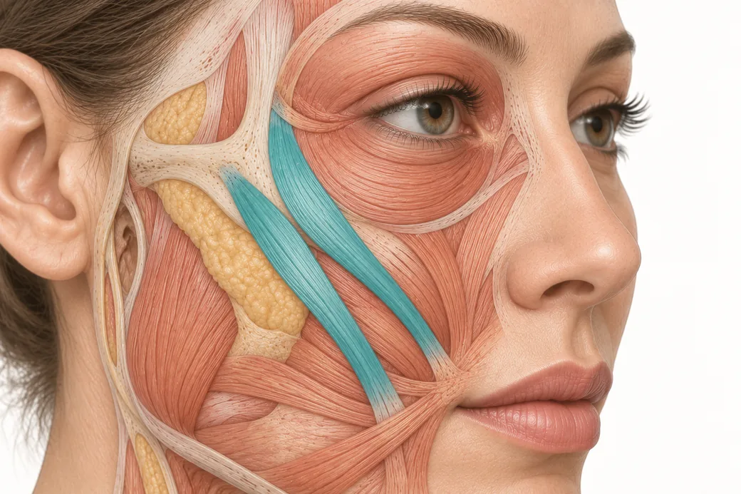 Clinical GuideThe Crow's Feet Trap — How Chasing Lateral Cheek Lines Reaches the ZygomaticusA zygomaticus botox complication after crow's feet treatment has one usual cause — chasing lateral cheek lines too low and too deep. Th
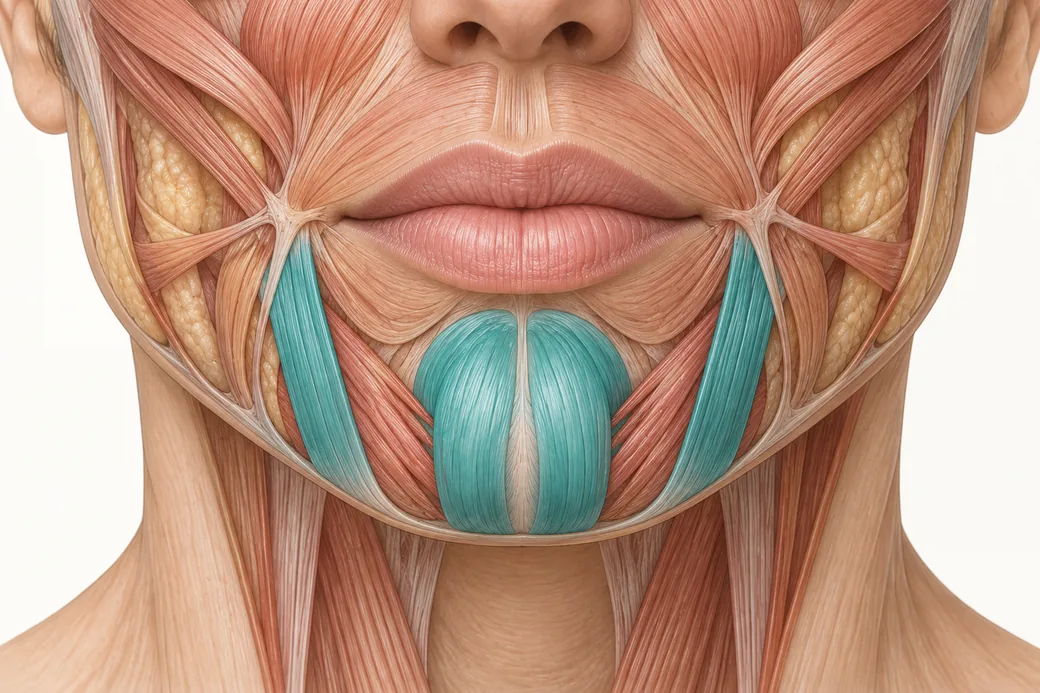 Clinical GuideDepressor Anguli Oris, DLI and Mentalis — Three Overlapping Muscles, Millimetres ApartDepressor anguli oris anatomy is a depth problem, not a map problem. The DAO, DLI and mentalis overlap in layers, and confusing them al
Train with Empire
This guide is clinical education. The technique behind it is taught hands-on, on live patients, with faculty beside you.
Explore Botox Training & Certification →Disclaimer
This article reflects the clinical opinions and experience of Melissa Pulcini-Buttine, PA, an independent faculty member contributing to Empire Medical Training's curriculum. The views expressed are the author's own and do not necessarily represent those of Empire Medical Training.
It is professional education, not medical advice, and is no substitute for hands-on training or independent clinical judgment. Licensed clinicians remain responsible for their own patient selection, technique and outcomes, for verifying current product labelling, and for practising within their scope and applicable law. Empire Medical Training accepts no liability for reliance on this content.
 Claim Your Prize!