







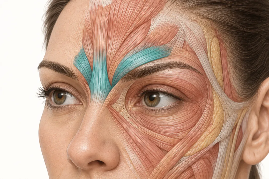
Corrugator muscle variation is the reason two competent injectors treat the same glabella with visibly different patterns, and it is the anatomical answer to one of the most awkward sentences a patient can say in a consultation: my last injector did it differently. Melissa Pulcini-Buttine, PA, who has taught anatomy and physiology for roughly fourteen years, addresses it directly in teaching because her own pattern changes from patient to patient:
"There's variation between people's muscles. That's why you have to have your patient make the movements. Because in some patients the corrugators go more horizontal, and in some patients it goes more vertical. And the only way you could see that is if you have them move."
"So sometimes my injection pattern might be like this, where the tail is more vertical. So it's not that it's wrong. If you see me injecting different patients and you say, well Melissa, one time you went more horizontal, one time you went more vertical — that's because I'm assessing the movement of the muscle."
This resource is about that assessment: what actually varies, how to read it on a moving face, and how to have the conversation with a patient who has interpreted variation as inconsistency. It is not a depth guide — the medial head and lateral tail require different depths for reasons covered in a companion resource in this cluster, and that logic sits alongside this one rather than inside it.
What the corrugator is doing
The corrugator supercilii is one of the brow depressors of the glabellar complex, working with the procerus to pull the brow down and medially in the expression the patient recognises as frowning. Melissa describes it functionally: "The procerus and the corrugators are natural depressors. When you're frowning and you're angry, they're pulling down."
Anatomically the muscle is usually described in two parts — a medial head and a lateral tail — and the tail is the part that varies. It is the tail that determines how far laterally the muscle's influence extends, how much of the brow it depresses, and crucially how close its fibres run to the frontalis fibres above them.
That last relationship is the one that makes orientation clinically consequential rather than merely interesting, and Melissa states the constraint that follows: "We're staying medial. We don't want to go lateral because we don't want to hit the fibres of the frontalis muscle." Where "lateral" begins is not a fixed distance. It is a function of where this patient's tail actually runs.
What varies, and by how much
Three separate dimensions vary between patients, and they vary independently.
Orientation. The tail may run close to horizontal — a shallow, laterally directed sweep along the brow — or more obliquely upward, producing what Melissa describes as a more vertical tail. The resulting glabellar lines differ accordingly, because a crease forms perpendicular to the fibre direction that produced it. A patient with vertical glabellar lines and a patient whose frown produces an oblique crease running up toward the medial forehead are showing you two different fibre orientations.
Extent. How far laterally the tail reaches determines how much of the brow it depresses and where its territory ends. This is what decides how far lateral a treatment can reasonably go before it stops being corrugator treatment and becomes frontalis treatment.
Size and depth. High-frequency ultrasound of the upper facial muscles in 127 healthy adults measured thickness and depth from the epidermis for the frontalis, procerus, corrugator supercilii and orbicularis oculi, and found relative side-to-side asymmetry coefficients reaching 40 percent overall, with individual muscles including the corrugator reaching 50 percent (Toxins 2025;17(12):595). BMI correlated moderately with the depth of the forehead muscles; men had thicker frontalis and procerus. The right corrugator and the left corrugator in the same patient are frequently not the same muscle in thickness or in depth below the skin.
There is a fourth variable that is easy to overlook: strength asymmetry. Melissa treats it as routine rather than exceptional. "Sometimes they're asymmetrical and this side pulls down more than this side. And I can compensate for that with the amount of injections that I do, or the units." Reading which side pulls harder is only possible under animation, and it is a separate finding from which side's muscle is larger.
Why a standard glabellar pattern is a starting point rather than an answer
The conventional glabellar approach is a five-point pattern: the head and tail of each corrugator plus the procerus. It is widely taught, it is what the registration trials used, and it is a perfectly reasonable default for a first treatment on an unfamiliar face.
Its limitation is structural. A five-point pattern encodes an assumption about where the tails run. Applied to a patient whose tails run more obliquely, the lateral points sit off the muscle — either too low, leaving the tail working, or too high, catching the frontalis fibres that Melissa is explicitly steering away from. The published critique of standardised approaches makes the same point more generally: on-label patterns were designed around clinical trials for registration purposes rather than real-world practice, and applying a single pattern to every patient fails to address individual anatomical variation (Aesthetic Surgery Journal Open Forum 2025;7:ojaf032). Among the documented consequences are the sharply upturned lateral brow and eyebrow splaying — both of which are shape errors traceable to treating the wrong part of the pair.
There is a second reason the tails deserve individual reading. Micro-innervation mapping of the upper facial muscles found that neuromuscular junctions are not uniformly distributed but cluster in muscle-specific motor zones: in the corrugator supercilii they concentrate predominantly within the medial portion and at greater depth, while in the orbicularis oculi they are diffusely distributed and in the frontalis they concentrate in the mid-to-upper belly along the deep fascial surface (Journal of Cosmetic Dermatology 2026;25(5):e70921). The authors are careful to describe these as probabilistic fields rather than coordinates. The practical reading is that the medial corrugator is comparatively forgiving of small placement variation because its motor territory is concentrated, and the tail — which is exactly where orientation varies most — is the part where getting the fibre direction right matters most.
Reading orientation at the chairside
The examination is short and it is the same one Melissa uses for everything else in this cluster.
Ask for the specific expression. "You're going to have your patient make angry face and frown." Not a general animation — a frown, held, repeated.
Watch the crease, then infer the fibre. Creases form perpendicular to the direction of pull. Vertical glabellar lines indicate fibres running roughly horizontally across them. An oblique crease running superolaterally indicates a more obliquely oriented tail.
Palpate during contraction. The contracted belly is findable under the fingertip in most patients, and the direction in which it firms is more informative than its position at rest.
Compare sides explicitly. Do not assume the orientation you identified on the right applies on the left. Note which side depresses further and faster.
Mark after the frown, not before. A mark placed before animation records the pattern you expected.
The "my last injector did it differently" conversation
This is where the anatomy pays for itself, because the patient is asking a reasonable question and the wrong answer damages two clinicians at once.
Dismissing the previous injector is the tempting move and the worst one. It teaches the patient that injectors disagree about basic facts, which makes every future recommendation — including yours — negotiable. Claiming there is one correct pattern is also false, and it sets you up badly when your own pattern changes at the next visit.
The accurate answer is short and it is the one Melissa gives her own trainees: patterns follow the muscle, the muscle differs between people and between sides, and the way it is identified is by watching the face move. "It's not that the injector is wrong. It's just we're watching the movement, and their muscle may be more horizontal and others might be more vertical."
Two things make that answer land. First, demonstrate it — have the patient frown while you describe what you are seeing, in the mirror. A patient who has watched you read their own anatomy stops experiencing variation as inconsistency. Second, say in advance that your own pattern may change at the next visit, and why: because muscles change with treatment history, age and residual toxin. That single sentence at visit one prevents the same conversation at visit three.
For the broader regional context, Empire's botox face chart and botox injection sites overviews are useful reference for orienting a patient during that conversation, and the distinction between a crease that still needs a muscle and one that has become fixed is covered in dynamic vs static wrinkles.
What changes at the chairside
- Treat the standard five-point glabellar pattern as a default, not a finding. Confirm the tails before you use it.
- Read orientation on every new patient and re-read it annually. It is a finding, not a fixed attribute.
- Record the orientation and the dominant side in the note. "Right tail oblique, left more horizontal, right depresses harder" is the note that makes your next visit shorter and better.
- Let the tail's actual course define where lateral begins. The instruction to stay medial is a relationship, not a measurement.
- Warn the patient in advance that patterns vary between visits and between clinicians, and explain why once rather than defending it later.
Reading muscle orientation from movement is a trained skill and it is learned fastest where the structure can be seen after it has been injected. Empire's anatomical based aesthetics training and special anatomical cadaver aesthetics training are built around exactly that sequence.
The clinical approach above reflects Melissa Pulcini-Buttine's practice as taught in Empire Medical Training's hands-on curriculum. Technique is learned under supervision; this article is educational and is not a substitute for training.
Melissa Pulcini-Buttine, PA is a physician assistant of two decades and has been a professor of anatomy and physiology for approximately fourteen years. She is a faculty member at Empire Medical Training and the founder of an aesthetics practice in Greenwich, Connecticut.
Related guides in this cluster
Part of Facial Muscle Anatomy for Neurotoxin.
 Clinical GuideMost Neurotoxin Complications Are Diffusion, Not Misplacement — Rethinking Root CauseMost neurotoxin complications come from diffusion into an adjacent muscle, not from injecting the wrong site. Here is how to tell which
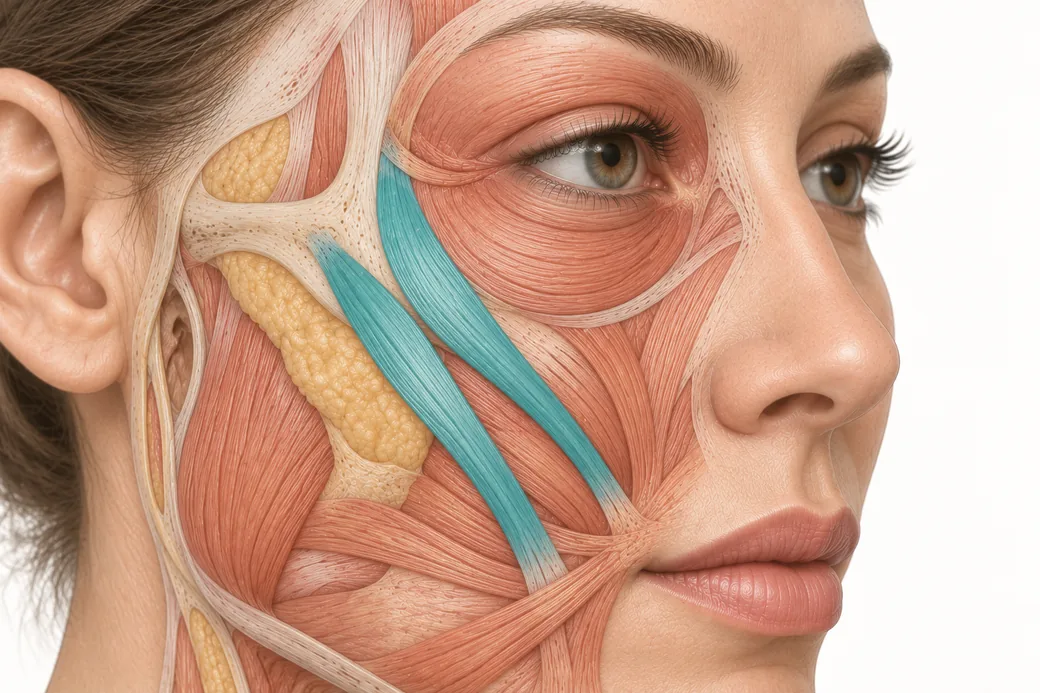 Clinical GuideThe Crow's Feet Trap — How Chasing Lateral Cheek Lines Reaches the ZygomaticusA zygomaticus botox complication after crow's feet treatment has one usual cause — chasing lateral cheek lines too low and too deep. Th
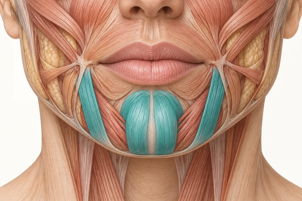 Clinical GuideDepressor Anguli Oris, DLI and Mentalis — Three Overlapping Muscles, Millimetres ApartDepressor anguli oris anatomy is a depth problem, not a map problem. The DAO, DLI and mentalis overlap in layers, and confusing them al
Train with Empire
This guide is clinical education. The technique behind it is taught hands-on, on live patients, with faculty beside you.
Explore Botox Training & Certification →Disclaimer
This article reflects the clinical opinions and experience of Melissa Pulcini-Buttine, PA, an independent faculty member contributing to Empire Medical Training's curriculum. The views expressed are the author's own and do not necessarily represent those of Empire Medical Training.
It is professional education, not medical advice, and is no substitute for hands-on training or independent clinical judgment. Licensed clinicians remain responsible for their own patient selection, technique and outcomes, for verifying current product labelling, and for practising within their scope and applicable law. Empire Medical Training accepts no liability for reliance on this content.
 Claim Your Prize!