







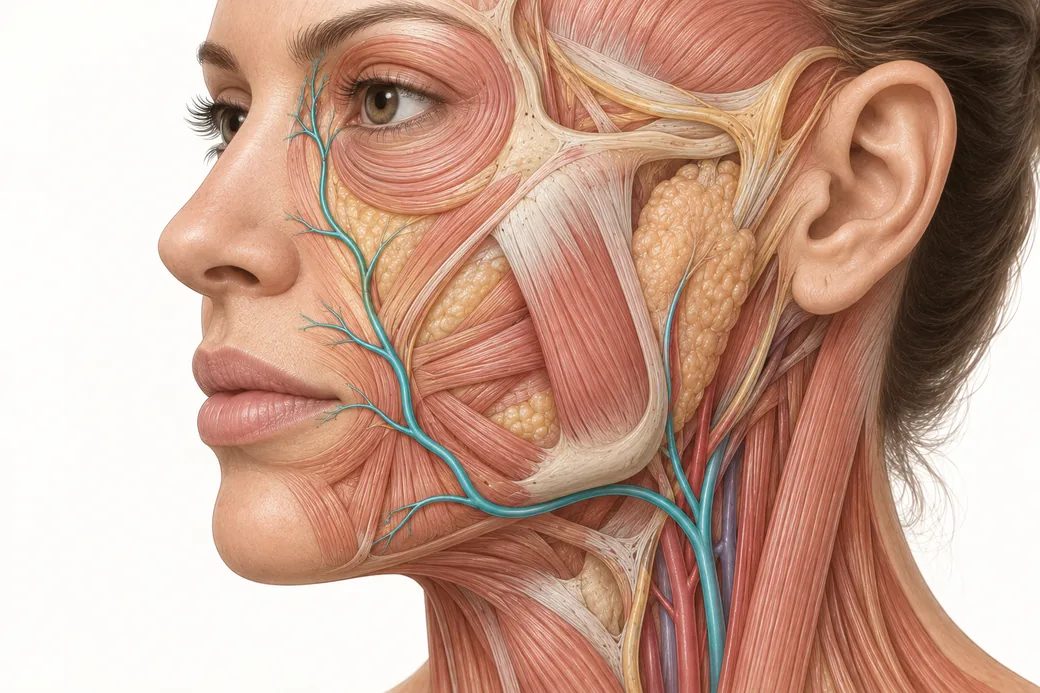
The trigeminal nerve map is the planning layer that sits in front of every facial block you will ever place. Not a memory test — a planning tool. If you know which division owns the territory you are about to treat, you know which foramen to aim at, how many injections the plan requires, and, just as importantly, which parts of the face are going to stay stubbornly awake no matter what you do. Injectors who skip this step end up placing blocks that do not cover the field, then concluding that blocks do not work.
There are two anatomical maps an injector needs and they answer opposite questions. The vascular map — covered in Melissa Pulcini-Buttine's facial arterial anatomy work — tells you where not to put a needle, and what happens if you do. The sensory map tells you exactly where to put one. They are complementary and they intersect at a specific and important place: the three foramina you target for facial blocks are neurovascular bundles, not isolated nerves. Treat the two maps as a set.
The structure before the territories
The trigeminal nerve is cranial nerve V. It is the principal sensory nerve of the face, and it also carries motor supply to the muscles of mastication — but only in its third division. That asymmetry matters more than it sounds, because it is the reason a V3 injection can affect function in a way a V1 or V2 injection cannot.
The three divisions arise from the trigeminal ganglion, and each leaves the skull by a different route:
| Division | Exits the skull via | Carries |
|---|---|---|
| V1 — ophthalmic | Superior orbital fissure | Sensory only |
| V2 — maxillary | Foramen rotundum, then the pterygopalatine fossa | Sensory only |
| V3 — mandibular | Foramen ovale | Sensory and motor to the muscles of mastication |
For office purposes, what you actually care about is the terminal branch and the surface foramen it emerges from — the point where the nerve becomes reachable with a 30-gauge needle and a couple of millilitres of lidocaine.
V1 — ophthalmic: the upper third, and the scalp all the way back
This is the division most commonly described incorrectly, so it is worth being exact.
Territory. The forehead. The anterior scalp, continuing posteriorly to the vertex of the skull. The upper eyelid and the conjunctiva and cornea. The root, dorsum and tip of the nose. The frontal sinus and part of the nasal mucosa.
The detail people get wrong is the scalp. Coverage to the crown belongs to V1 — specifically to the supraorbital and supratrochlear nerves, which run vertically from the superior orbital rim up over the frontalis and continue back across the scalp. It does not belong to V2. This is not a trivia point: it is the reason the anterior two-thirds of a scalp block is achieved at the eyebrow, and it is the reason a midface block will do nothing for a patient having a forehead procedure.
Branches that matter to an injector:
- Frontal nerve, which divides into the supraorbital and supratrochlear nerves. These are the two you block, and together they own the forehead and the scalp to the vertex.
- Infratrochlear nerve, supplying the medial canthus, the lacrimal sac region and the side of the nasal root.
- Nasociliary → anterior ethmoidal → external nasal branch, which emerges at the junction of the nasal bone and the upper lateral cartilage and supplies the tip of the nose. This is the single most useful obscure fact on this page. Nasal tip sensation is V1, not V2. An infraorbital block that numbs the ala beautifully will leave the tip sensate, and a patient having non-surgical rhinoplasty will tell you so.
What V1 buys you clinically: glabellar complex work, forehead toxin, forehead and temple filler, hairline and anterior scalp PRP or microneedling, brow work, and the upper portion of a nasal procedure.
V2 — maxillary: the midface
Territory. Lower eyelid. The cheek and midface. The lateral nose and ala. The upper lip. The upper teeth and upper gingiva. The maxillary sinus and most of the hard palate.
Branches that matter:
- Infraorbital nerve, the terminal branch, emerging from the infraorbital foramen and immediately fanning into inferior palpebral, lateral nasal and superior labial branches. This one branch is the workhorse of midface anesthesia.
- Zygomaticofacial, supplying skin over the body of the zygoma.
- Zygomaticotemporal, supplying the skin of the temple — a useful contributor to a scalp block and easy to forget.
- Superior alveolar nerves, to the upper dentition.
What V2 buys you clinically: tear trough and infraorbital hollow, cheek and malar filler, nasolabial folds, the ala and lateral nose, and — the big one — the upper lip. One infraorbital block per side covers most of what an injector does in the midface.
Where V2 stops. It does not reach the forehead. It does not reach the scalp. It does not reach the nasal tip, which is V1, or the lower lip and chin, which are V3.
V3 — mandibular: the lower third, and the only motor division
Territory. Lower lip. Chin. Lower teeth and lower gingiva. The buccal mucosa. General sensation to the anterior two-thirds of the tongue. The temple and preauricular skin via the auriculotemporal nerve. Part of the external auditory meatus and the tympanic membrane.
Branches that matter:
- Inferior alveolar nerve → mental nerve, emerging from the mental foramen. The mental nerve is the one you block for lower lip, chin and marionette work.
- Buccal nerve (sensory to the cheek mucosa and a patch of skin overlying buccinator — distinct from the buccal branch of the facial nerve, which is motor; do not confuse them).
- Auriculotemporal nerve, which passes just posterior to the temporomandibular joint and ascends with the superficial temporal artery. It supplies the temple and preauricular region and is a component of a complete scalp block.
- Lingual nerve, to the tongue.
Motor. V3 supplies masseter, temporalis and the pterygoids, plus mylohyoid, anterior digastric, tensor tympani and tensor veli palatini. This is the division that intersects with masseter neurotoxin and bruxism work — a relationship covered in our pieces on managing bruxism and Botox for jaw clenching.
What V3 buys you clinically: lower lip, chin projection and chin dimpling, marionette lines, prejowl sulcus, and the mandibular border.
The part everyone gets wrong: what is not trigeminal
The single most useful thing on this map is its edge.
The angle of the mandible is not trigeminal. The skin over the angle of the jaw, the parotid region, and the lower preauricular area are supplied by the greater auricular nerve, which is a branch of the cervical plexus arising from C2 and C3. No amount of V3 block reaches it.
The posterior scalp is not trigeminal. Behind the vertex, sensation belongs to the greater occipital nerve (C2, from the dorsal ramus) and the lesser occipital nerve (C2–C3, cervical plexus).
The neck is not trigeminal. Transverse cervical and supraclavicular nerves, again cervical plexus.
So the trigeminal territory has a real posterior and inferior boundary, running roughly from the vertex down in front of the ear to the angle of the mandible. Everything behind and below that line is cervical.
Two consequences follow, and both are practical.
First, you should expect areas of retained sensation and you should say so out loud before you start. A patient who has been told "I am going to numb your face" and who then feels the jawline sharply concludes the block failed. A patient who has been told "the jaw angle is supplied by a different nerve from the neck and will stay awake" concludes that you knew what you were doing. Same sensation, completely different experience.
Second, a genuinely complete scalp block is not a trigeminal block. It is a trigeminal block plus a cervical one — V1 anteriorly, zygomaticotemporal and auriculotemporal laterally, and the greater and lesser occipital nerves posteriorly.
The planning table
This is the map as an injector actually uses it — target first, nerve second.
| Treatment area | Territory | Division | Where you would block |
|---|---|---|---|
| Glabella, forehead, brow | Forehead | V1 | Supraorbital + supratrochlear, at the notch |
| Anterior scalp to vertex | Scalp | V1 | Supraorbital + supratrochlear |
| Nasal tip | Nasal tip | V1 (external nasal) | Not reached by an infraorbital block |
| Temple | Temple | V2 (zygomaticotemporal) + V3 (auriculotemporal) | Lateral orbital rim / preauricular |
| Tear trough, cheek, malar | Midface | V2 | Infraorbital foramen |
| Ala, lateral nose | Lateral nose | V2 | Infraorbital |
| Upper lip, philtrum | Upper lip | V2 | Infraorbital |
| Lower lip | Lower lip | V3 | Mental foramen |
| Chin, marionettes, prejowl | Chin | V3 | Mental foramen |
| Angle of jaw, parotid region | Cervical | Greater auricular, C2–C3 | Not trigeminal — separate block |
| Posterior scalp / occiput | Cervical | Greater + lesser occipital | Not trigeminal — separate block |
Read down the middle column when a patient asks why one area went numb and another did not.
Where the sensory map meets the vascular map
The three surface foramina you target are not nerve-only structures. Each transmits a neurovascular bundle:
- Supraorbital foramen / notch — supraorbital nerve with the supraorbital artery and vein.
- Infraorbital foramen — infraorbital nerve with the infraorbital artery and vein.
- Mental foramen — mental nerve with the mental artery and vein.
That is precisely why a block site is also a bruising site and an intravascular-injection site, and it is why the safety principles for these blocks are aspiration, small volumes, and slow delivery rather than confident speed. The nerve you are aiming for is travelling with an artery that is a branch of the same system a filler embolus would travel in. If you have not read the arterial map recently, read it alongside this one.
The variability matters too. The supraorbital notch is palpable in most patients but can be a closed foramen sited slightly higher. Infraorbital foramen position varies by roughly a finger's breadth below the orbital rim and is not perfectly on the mid-pupillary line in every face. The three foramina do tend to fall roughly in a vertical line with one another, which is the most useful single landmark heuristic on the face — but treat it as an orientation, not a coordinate.
What a sensory block does and does not abolish
A successful field block removes sharp pain from its territory. It does not remove everything, and the gap between those two statements is where patient complaints live.
Patients under a good infraorbital block will still report pressure, stretch and movement. Large-diameter fibres carrying pressure and proprioception are more resistant to local anesthetic than the small fibres carrying sharp pain, so differential blockade leaves the patient aware of what you are doing without it hurting. Cannula passage under a block feels like a firm push. Bolus placement feels like fullness.
Tell them this in advance in one sentence: "You will feel me pushing and moving things — you should not feel anything sharp." That sentence converts an alarming sensation into an expected one.
A sensory block also does not prevent bruising, does not reduce vascular risk, and does not compensate for imprecise depth. Comfort and safety are separate systems that happen to share an anatomy.
What to do with this on Monday
Before your next block, answer three questions in order. Which division owns the field I am treating? Which foramen does that division reach the surface through? Which parts of my treatment area fall outside trigeminal territory entirely, so that I can warn the patient now rather than explain later?
That is the whole planning layer. The technique for actually placing the four office blocks — supraorbital and supratrochlear, infraorbital, mental, and the greater auricular exception — is covered separately in this cluster, along with the maximum-dose calculation that has to precede any multi-site block.
Injectors who want this anatomy in three dimensions rather than in a table will get more out of Anatomical Based Aesthetics Training and Special Anatomical Cadaver Aesthetics Training than out of any amount of reading; the regional applications show up in Master Eye & Nose Injection Training and Complete Dermal Filler Training.
This material reflects Dr. Jennifer Thomas-Goering's clinical practice as taught in Empire Medical Training's hands-on curriculum. Technique is learned under supervision; this article is educational and is not a substitute for training.
Dr. Jennifer Thomas-Goering, DO, MBA is a board-certified anesthesiologist, clinical lead instructor and executive committee member at Empire Medical Training, and founder of an aesthetics practice in Ann Arbor, Michigan.
Related guides in this cluster
Part of Injectable Anesthesia and Patient Comfort.
 Clinical GuideBenzodiazepine or Clonidine? Choosing an Oral Anxiolytic for the Long Aesthetic AppointmentBenzodiazepine vs clonidine for pre-procedure anxiolysis — the mechanisms, the respiratory and scheduling differences, and the screenin
 Clinical GuideSublingual Ketamine in the Aesthetic Suite: Where It Fits, and What It Costs YouSublingual ketamine is the wrong tool for toxin and the right one for higher-discomfort work. The pharmacology, the FDA position, and t
 Clinical GuideNitrous Oxide in the Aesthetic Practice: Self-Titration, Contraindications and Same-Day DrivingNitrous oxide in aesthetic practice is the one non-controlled, self-limiting option. Titration range, contraindications, and the equipm
Train with Empire
This guide is clinical education. The technique behind it is taught hands-on, on live patients, with faculty beside you.
Explore Complete Dermal Filler Training →Disclaimer
This article reflects the clinical opinions and experience of Dr. Jennifer Thomas-Goering, DO, MBA, an independent faculty member contributing to Empire Medical Training's curriculum. The views expressed are the author's own and do not necessarily represent those of Empire Medical Training.
It is professional education, not medical advice, and is no substitute for hands-on training or independent clinical judgment. Licensed clinicians remain responsible for their own patient selection, technique and outcomes, for verifying current product labelling, and for practising within their scope and applicable law. Empire Medical Training accepts no liability for reliance on this content.
 Claim Your Prize!