







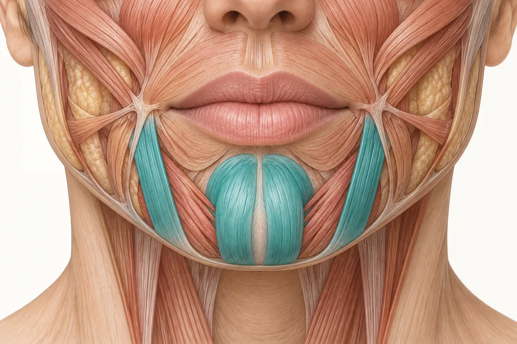
DAO neurotoxin concentration is the variable most injectors adjust last and should adjust first. The depressor anguli oris is a legitimate and common neurotoxin target. The depressor labii inferioris, which lies immediately medial to it and frequently overlaps it, is not. The distance between a good result and a visible complication in this corridor is measured in single millimetres — and at those distances, how much fluid you deposit matters as much as where you deposit it.
Dr. Chris Croley, Chief Medical Officer at Empire Medical Training, frames the problem directly in Empire's reconstitution curriculum:
"If we're injecting any of our neurotoxins around the mouth, those muscles are really close. So we look at things like the depressor anguli oris — that's here. Right next to this we have our depressor labii inferioris. One of these is a desired treatment for neurotoxin. The other is not. So you can see even a small movement of placement of my toxin or my syringe, or if I inject a large volume into this area, it can diffuse or move over into that area. So in those situations we'd like to use something with a very tight concentration."
This article is the dilution answer to that problem. The placement answer — landmarks, depth, entry angle, patient positioning — is covered separately in Empire's curriculum and in a companion piece from a different faculty session. The two answers are complementary and neither substitutes for the other.
The anatomy that sets the tolerance
The DAO is a thin, superficial, triangular strap arising from the oblique line of the mandible and converging on the modiolus at the corner of the mouth. Relaxing it allows the modiolus to reposition slightly upward, which is why it is targeted for the downturned mouth corner.
Contemporary ultrasound work has replaced a lot of textbook estimation with measured values, and the numbers explain why this corridor is unforgiving.
Depth. Alfertshofer and colleagues, publishing in Aesthetic Surgery Journal in 2024, imaged 80 DAO muscles in 40 toxin-naïve volunteers. Mean distance from skin surface to muscle surface was 4.4 mm, greater in males and increasing with BMI. Mean DAO thickness was 3.5 mm, ranging 2.8 to 4.8 mm, with thinner muscles in females. The authors calculated the most favourable intramuscular injection depth at 6.1 mm.
Proximity and overlap. Zhang and colleagues, in the same journal in 2024, imaged 41 patients and quantified the relationship between DAO and DLI at the labiomandibular fold. Skin-to-muscle depths averaged 5.26, 5.61 and 8.42 mm from the medial to lateral border. Critically, the overlapping length of DAO and depressor labii inferioris increased from 4.74 mm in zone 1 to 9.68 mm in zone 2 and 14.54 mm in zone 3. The medial border of the DAO sat 4.33, 6.12 and 8.90 mm medial to the labiomandibular fold across those zones — and in nearly one third of patients, no DAO fibres were observed in zone 1 or zone 2 at all.
Neighbours on the other side. Yi and colleagues, in Anatomy and Cell Biology in 2023, describe the full problem: the DAO's medial border overlaps the depressor labii inferioris, while its lateral border is adjacent to risorius, zygomaticus major and platysma. Their stated aim was to reduce both dose and number of injection points precisely because of that crowding.
Put those findings together and the clinical picture is stark. You are aiming at a muscle roughly three and a half millimetres thick, lying about four to five millimetres below the skin, which overlaps a muscle you must not weaken by up to fourteen millimetres, and which in a meaningful minority of patients is not where you expect it to be.
What happens when the field is too wide
The DLI depresses and everts the lower lip. Partial chemodenervation of it produces a specific and recognisable presentation: asymmetry of the lower lip on animated speech and smiling, with the affected side failing to depress. Patients describe difficulty pronouncing certain consonants, biting the lower lip, and a smile that "doesn't work on one side." It is highly visible in conversation, it does not respond to anything but time, and it persists for the duration of the toxin's effect.
The related error — spread into risorius, zygomaticus major or platysma laterally — produces its own asymmetries. Yi and colleagues note explicitly that a lack of knowledge of DAO anatomy and of toxin properties can lead to side effects such as asymmetrical smiles.
This is the consequence weighting that should drive your concentration choice. The probability of overreach in this corridor may be modest; the visibility and duration of the result are not.
The concentration answer
Croley's instruction is to raise concentration so that the intended dose occupies the smallest practical volume:
"Maybe we would use something like a one millilitre reconstitution. So for me to deliver two units I would only go to this part of the syringe. Tiny, tiny dose. So it doesn't go over here."
Work the arithmetic across the preparations a practice realistically holds, for a 2 Unit per side DAO dose:
| Reconstitution of a 100 Unit vial | Concentration | Volume for 2 Units | Volume for 3 Units |
|---|---|---|---|
| 1 mL | 10 Units/0.1 mL | 0.02 mL | 0.03 mL |
| 1.25 mL | 8 Units/0.1 mL | 0.025 mL | 0.0375 mL |
| 2 mL | 5 Units/0.1 mL | 0.04 mL | 0.06 mL |
| 2.5 mL | 4 Units/0.1 mL | 0.05 mL | 0.075 mL |
| 4 mL | 2.5 Units/0.1 mL | 0.08 mL | 0.12 mL |
At the routine 2.5 mL working concentration, a 2 Unit DAO dose deposits 0.05 mL. At a 1 mL reconstitution the same dose deposits 0.02 mL — 60% less fluid into a corridor where the protected muscle may overlap the target by several millimetres. At a 4 mL dilute preparation it deposits 0.08 mL, four times the concentrated volume, into the same space.
The relevance of that volume difference is not theoretical. Hsu, Dover and Arndt demonstrated in Archives of Dermatology in 2004 that an identical 5 Unit dose delivered in 0.25 mL rather than 0.05 mL produced a roughly 50% larger area of effect on the forehead. Volume moves the boundary of the field. In the perioral corridor, the boundary is the whole problem.
These figures reflect Dr. Croley's clinical practice as taught in Empire Medical Training's hands-on curriculum. Technique is learned under supervision; this article is educational and is not a substitute for training.
The trade-offs a concentrated preparation brings with it
Raising concentration solves the spread problem and creates three smaller ones. All are manageable; none should be a surprise.
You cannot read 0.02 mL on every syringe. A 0.02 mL aliquot is one fifth of the smallest labelled increment on a standard 1 mL syringe. This is where syringe selection stops being a preference and becomes part of the dose. Empire's companion resource on insulin, 0.5 mL and 1 mL syringe graduations covers the mechanics; the short version is that a syringe whose graduations you cannot resolve at your working volume is the wrong syringe for this treatment.
Dead space costs more. Residual fluid in the needle hub is a fixed volume and a variable number of units. At 10 Units per 0.1 mL, a 0.05 mL residue is 5 Units of product discarded — more than an entire bilateral DAO treatment.
Small errors are amplified. At 10 Units per 0.1 mL, a 0.01 mL overdraw is 1 Unit. On a 2 Unit target that is a 50% dosing error. Concentrated preparations reduce spread risk and increase measurement risk, which is why they belong with a syringe and a technique matched to them rather than with the barrel that happens to be on the tray.
Concentration is one control, not the only one
Nothing here replaces placement. The published anatomy makes that unambiguous: Zhang and colleagues recommend injecting at the middle and lower thirds of the labiomandibular fold at a 4 to 5 mm depth, and note that nearly a third of patients have no DAO fibres in the upper zone at all. Yi and colleagues advocate reducing both dose and injection point count. Alfertshofer and colleagues calculate 6.1 mm as the favourable intramuscular depth and demonstrate that sex and BMI move it.
Three controls act together, and concentration is the one this cluster owns:
Dose. Fewer units means a smaller field at any concentration. Conservative starting doses with a two-week review are appropriate in a first-time perioral patient.
Placement and depth. Position relative to the labiomandibular fold and the mandibular border, and depth within the muscle rather than superficial or deep to it. Owned by Empire's anatomy curriculum and by the companion placement resource.
Concentration. The volume the dose occupies once it is in tissue. Owned here.
An injector who optimises placement while injecting 0.08 mL of a dilute preparation has undone the precision they worked for. An injector who uses a concentrated preparation but places it laterally at the wrong depth has not been rescued by their arithmetic. The corridor demands both.
Injectors who want to work through perioral anatomy with supervision can do so in Empire's Anatomical Based Aesthetics Training and Special Anatomical Cadaver Aesthetics Training, and the vial and dosing work in Cosmetic Neurotoxins Training. Empire's material on chin dimpling and chin injection sites covers adjacent lower-face targets that share this corridor.
Related guides in this cluster
Part of Neurotoxin Reconstitution and Dosing.
 Clinical GuideOn-Label Reconstitution Across the US Neurotoxins — A Reference Table for InjectorsA complete on-label neurotoxin reconstitution chart for Botox, Dysport, Xeomin, Jeuveau and Daxxify — vial sizes, diluent volumes and u
 Clinical GuideWhy a Unit of Botox Is Not a Unit of DysportBotox units vs Dysport units are not the same measure. Units are defined by each manufacturer's own potency assay — here is why no conv
 Clinical GuideBacteriostatic vs Preservative-Free Saline — What the Benzyl Alcohol Actually DoesBacteriostatic saline for neurotoxin reconstitution is off-label but well studied. What benzyl alcohol does to injection pain, potency
Train with Empire
This guide is clinical education. The technique behind it is taught hands-on, on live patients, with faculty beside you.
Explore Botox Training & Certification →Disclaimer
This article reflects the clinical opinions and experience of Dr. Chris Croley, Chief Medical Officer, Empire Medical Training, an independent faculty member contributing to Empire Medical Training's curriculum. The views expressed are the author's own and do not necessarily represent those of Empire Medical Training.
It is professional education, not medical advice, and is no substitute for hands-on training or independent clinical judgment. Licensed clinicians remain responsible for their own patient selection, technique and outcomes, for verifying current product labelling, and for practising within their scope and applicable law. Empire Medical Training accepts no liability for reliance on this content.
 Claim Your Prize!