







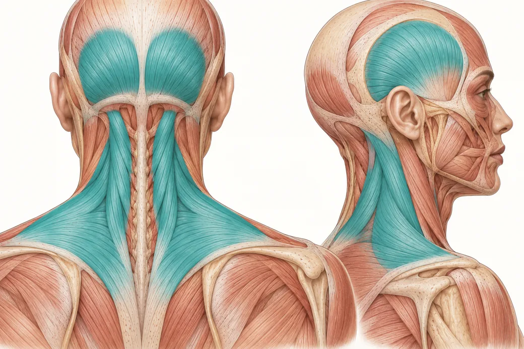
Trapezius and occipitalis botox injections, along with the cervical paraspinal group, are the three fields in the chronic migraine protocol that no aesthetic training programme has any reason to teach you. They are also, between them, more than half the injection sites in the approved regimen. That arithmetic is the whole problem: an injector who treats only the fields they already know is not delivering a reduced version of the protocol, they are delivering a different treatment with the same name.
When we look at the anterior part of the face, the frontalis and corrugator region, those areas are fairly close to territory that is on-label and approved for migraine injection, and we treat them every day. But as we start to move posteriorly — the trapezius, the occipitalis — those are not areas we would typically treat for aesthetic purposes. So many of us in aesthetic practice start by treating the fields we are comfortable with, and if the patient is not getting a response, we should consider that in aesthetics we are often under-treating the on-label way this has traditionally been used.
This article is the anatomy and the rationale. It does not give per-site unit allocations; those belong in the BOTOX prescribing information, and they should be read there.
Why this is a training gap rather than a skills gap
Aesthetic injecting is anterior. The muscles you have spent your career learning — frontalis, corrugator, procerus, orbicularis oculi, the perioral group, masseter for some practices — are all visible from the front and all produce a visible surface effect. Empire's material on Botox injection sites and the Botox face chart maps that territory well.
The posterior fields produce no cosmetic effect anyone asks for. There is no aesthetic indication to relax the occipitalis. Nobody presents wanting a smoother cervical paraspinal group. So the anatomy is genuinely unfamiliar, the landmarks are palpated rather than observed, and the failure modes are ones an aesthetic injector has never had to think about — because in aesthetics the consequence of a misplaced injection is a brow that sits wrong for three months, and here it is a neck that will not hold the head up comfortably.
That is not a reason to avoid the territory. It is a reason to learn it properly before entering it.
Occipitalis
Attachments and course. The occipitalis arises by tendinous fibres from the lateral two-thirds of the highest nuchal line of the occipital bone and the adjacent region of the mastoid part of the temporal bone, and extends upwards and laterally to join the aponeurotic fascia.
Innervation. The posterior auricular branch of the facial nerve.
Vascular supply. Branches of the posterior auricular and occipital arteries.
Function — and the part that will feel familiar. Working with the frontalis, the occipitalis pulls the scalp backwards. The two should be considered a single functional muscle complex linked by fascia: the occipitalis is part of the fronto-occipital aponeurotic fascial system, and it is activated when frowning or elevating the eyebrows (Blumenfeld AM, Silberstein SD, Dodick DW, Aurora SK, Brin MF, Binder WJ. Insights into the Functional Anatomy Behind the PREEMPT Injection Paradigm: Guidance on Achieving Optimal Outcomes. Headache. 2017;57(5):766–777).
That last point is the hook I use when teaching this to aesthetic injectors. You already manipulate one end of this aponeurotic system every time you treat a frontalis. The occipitalis is the posterior half of a structure you know.
Landmarks. Three bony points define the field: the inion — the most posterior midline point of the occipital protuberance — the nuchal ridge, and the tip of the mastoid process behind the ear. The first injection site is found by placing the thumb on the inion and the index finger on the mastoid tip, halving the space between them, and injecting just above the nuchal ridge at that midpoint. The remaining sites step outwards and upwards from there toward the helix of the ear.
Technique principles. Injections are given at 45 degrees into the superficial aspect of the muscle, just below the dermis, with the needle angled upward away from the neck, and always above the nuchal ridge.
The failure mode. Patients may experience neck pain or weakness if injections are administered too low — below the nuchal ridge — and the injector must ensure these injections do not involve the suboccipital region. The sites also sit in close proximity to the greater and lesser occipital nerves, so patients should be told in advance that these injections may hurt more than the anterior ones. That single warning prevents a lot of patients from declining the posterior fields at cycle two.
Cervical paraspinal muscle group
Composition. This is not one muscle. The cervical paraspinal group is made up of multiple muscles including the trapezius, the splenius capitis and cervicis, and the semispinalis capitis.
- Splenius capitis ascends from the inferior half of the nuchal ligament and the spinous processes of the superior six thoracic vertebrae, attaching to the lateral aspect of the mastoid process and the lateral third of the superior nuchal line.
- Semispinalis capitis forms the longitudinal bulge in the back of the neck near the median plane and originates from the transverse processes of C4–T12.
Innervation — and why this field matters neurologically. The group is innervated by the posterior rami of the spinal nerves. The third occipital nerve traverses this group near the midline, and laterally the greater and lesser occipital nerves emerge through these muscles.
Vascular supply. Branches of the occipital artery.
Function. Support and stabilisation of the neck, plus rotation and extension of the head.
Technique principles. Injections go into the most superficial aspect of the muscle near the midline, with the needle angled at 45 degrees and superiorly. Position matters: the patient should be upright with the head in a neutral position, because a neck flexed too far forward turns a superficial injection into a deep one. Penetrating the fascia, which is variable in thickness, should be sufficient depth.
The failure mode, and the mental model that prevents it. Injections that are too low or too deep in this group can lead to muscle weakness and neck pain. The guidance that keeps you out of trouble is to think of the cervical paraspinals as suboccipital muscles, so that the sites cannot drift down the neck — and to visualise a horizontal line across the neck roughly two fingerbreadths below the occipital protuberance and stay above it. In general the injections should sit higher, just inferior to the nuchal ridge, where there is a thick fascial condensation that minimises the potential for exacerbated neck weakness. These injections occur in the hairline (Blumenfeld et al., 2017).
Assess before you treat: chronic migraine patients may arrive with preexisting neck pain or weakness, and you need to have documented it so that you are not later trying to distinguish a baseline complaint from a treatment effect.
Trapezius
Attachments. A large, flat, triangular, superficial muscle attaching proximally at the medial third of the superior nuchal line, the external occipital protuberance, the nuchal ligament and the spinous processes of C7–T12, and distally at the lateral third of the clavicle, the acromion and the spine of the scapula.
Innervation. The spinal accessory nerve (CN XI) supplies the motor fibres, with C3 and C4 spinal nerves carrying pain and proprioceptive fibres. The sensory rami of C2, C3 and C4 run across this muscle.
Vascular supply. The transverse cervical artery.
Function. Stabilisation and movement of the scapula, and support for the arm. That is the functional stake: this is not a cosmetic muscle whose weakness costs an expression, it is a muscle whose weakness costs shoulder function.
Landmarks. The field is the upper portion of the muscle between the inflection point of the neck — the "necklace line" — and the acromioclavicular joint, with sites placed at the midpoint of that span and at the midpoints between it and each end.
Technique principles. Injections belong in the supraclavicular portion, lateral to the neckline and medial to the deltoid and the acromioclavicular joint, delivered horizontally and superficially. Where skin or subcutaneous tissue is thick, deeper penetration may be needed simply to reach superficial muscle.
The failure modes, plural. Injecting too high or too deep may cause neck or shoulder weakness, as well as neck pain from compensatory muscle activity. Injecting too far laterally puts drug into the deltoid, which produces shoulder weakness. And patients with small frames may be predisposed to weakness after injection here, which should be assessed and discussed at baseline (Blumenfeld et al., 2017).
There is one further caution worth carrying. Where additional follow-the-pain units are permitted, the published guidance is that neck weakness and neck pain are associated with high doses into the trapezius, and that additional trapezius injections should therefore generally be avoided where possible even under a follow-the-pain strategy. Of the three posterior fields, this is the one where more is least likely to be better.
Why omitting these fields is not a dose reduction
The intuitive reading of skipping the posterior third is that you are giving a lower dose of the same treatment. The anatomy says otherwise.
The sensory nerve endings that this treatment acts on belong to neurons whose cell bodies sit in both the trigeminal ganglia and the cervical ganglia, and they are distributed throughout all of the injected muscles. Spinal nerves originating from the C2 and C3 dorsal root ganglia innervate the pericranial muscles and extend intracranially through cranial sutures, emissary canals and fissures — the anterior fields are where the trigeminal contribution is accessible, and the posterior fields are where the cervical contribution is (Burstein R, Blumenfeld AM, Silberstein SD, Manack Adams A, Brin MF. Mechanism of Action of OnabotulinumtoxinA in Chronic Migraine: A Narrative Review. Headache. 2020;60(7):1259–1272).
Omitting the posterior fields therefore does not scale the treatment down. It removes an entire anatomical input from a mechanism that depends on both. That is the anatomical form of the clinical observation: aesthetic-pattern injectors systematically under-treat the on-label protocol, and then conclude from a partial response that the patient is a poor responder.
The published guidance names the behaviour and its cost directly — lowering doses, avoiding muscles, or delaying repeat treatments may lead to suboptimal efficacy (Blumenfeld et al., 2017).
The adverse events you are avoiding are posterior-field adverse events
It is worth being explicit about why injectors omit these fields, because the reason is real.
In the chronic migraine trials behind the US label, adverse reactions reported in at least 2% of treated patients and more often than placebo included neck pain (9% versus 3%) and muscular weakness (4% versus <1%), alongside musculoskeletal stiffness, myalgia and eyelid ptosis. Looking specifically at injection-related events in the double-blind phases, neck pain was reported in 6.7%, muscular weakness in 5.5% and eyelid ptosis in 3.3% (Blumenfeld et al., 2017).
Neck pain and muscular weakness are the signature complaints of this treatment, and they come from these three fields. But read the technique sections above again: almost every mechanism by which they arise is a placement error. Too low, below the nuchal ridge. Too deep, because the neck was flexed. Too far lateral, into the deltoid. Too much into the trapezius.
The correct response to a posterior-field adverse event is better placement, not abandonment of the field. Omission converts a technique problem into an efficacy problem and hides it.
What to learn, and in what order
- Palpation first. Inion, nuchal ridge, mastoid tip, external occipital protuberance, necklace line, acromion. These are palpated landmarks on a patient sitting upright, and you should be able to find all of them with your eyes closed before you hold a syringe over them.
- Depth discipline second. Every posterior field in this protocol is a superficial injection with an upward or horizontal angle. The aesthetic instinct to seat a needle against periosteum is wrong here in all three fields.
- Positioning third. Upright, head neutral. Most depth errors in the cervical paraspinals are posture errors.
- Then the units, from the prescribing information, under supervision.
Rigorous regional anatomy is taught hands-on in Empire's Anatomical Based Aesthetics Training and, on cadaveric specimens, in Special Anatomical Cadaver Aesthetics Training. For injectors extending their work below the jawline more generally, the Neck & Hands Rejuvenation Master Course covers the region hands-on.
This guidance reflects Dr. Chris Croley's clinical practice as taught in Empire Medical Training's hands-on curriculum. Per-site dosing is deliberately not reproduced. Technique is learned under supervision; this article is educational and is not a substitute for training.
Related guides in this cluster
Part of Therapeutic Neurotoxin: Chronic Migraine.
 Clinical GuideChronic Migraine On-Label Criteria: Deciding Which Patients Actually QualifyChronic migraine criteria decide who you can treat on-label. The label wording, the ICHD-3 definition, the diary, and the documentation
 Clinical GuideConfirm the Diagnosis First: Secondary-Headache Red Flags Before You InjectSecondary headache red flags decide whether a migraine patient gets injected or referred. The SNNOOP10 framework, the numbers behind it
 Clinical GuideThe PREEMPT Protocol: Why Fixed-Site, Fixed-Dose Was the Trial Design, and What It Means in PracticeThe PREEMPT protocol is fixed-site, fixed-dose for a reason. What the trial design was, why the label is rigid, and where tailoring is
Train with Empire
This guide is clinical education. The technique behind it is taught hands-on, on live patients, with faculty beside you.
Explore Botox Training & Certification →Disclaimer
This article reflects the clinical opinions and experience of Dr. Chris Croley, Chief Medical Officer, Empire Medical Training, an independent faculty member contributing to Empire Medical Training's curriculum. The views expressed are the author's own and do not necessarily represent those of Empire Medical Training.
It is professional education, not medical advice, and is no substitute for hands-on training or independent clinical judgment. Licensed clinicians remain responsible for their own patient selection, technique and outcomes, for verifying current product labelling, and for practising within their scope and applicable law. Empire Medical Training accepts no liability for reliance on this content.
 Claim Your Prize!