







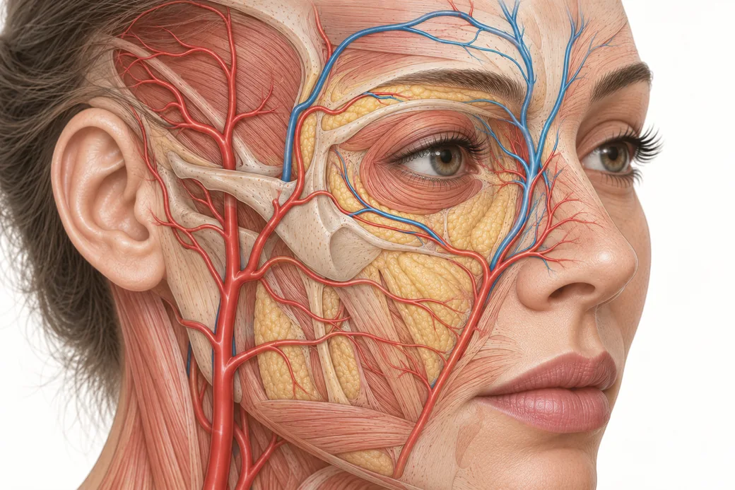
Most aesthetic practices need exactly four facial nerve blocks, and three of them are the same injection repeated at three different foramina. That is the good news. The less comfortable news is that these blocks have predictable coverage gaps, and an injector who does not know where the gaps are will spend the session apologising for an area that was never going to be numb.
This piece covers the three trigeminal field blocks an office realistically places — supraorbital with supratrochlear, infraorbital, and mental — plus the greater auricular block, which is the exception that makes the set make sense. It assumes you already know which division owns which territory; if not, read the trigeminal map first, because the map is the planning layer and this is the execution layer.
A note on framing before anything else. A block is a volume of local anesthetic placed near a nerve. That means every block is simultaneously a maximum-dose question and an intravascular-injection question, because all three facial foramina transmit a neurovascular bundle. Calculate your ceiling before you start; that calculation is covered separately in this cluster and it is not optional once you are placing four to six injections in one session.
The common technique, stated once
The three trigeminal blocks share a method. Learn it once.
- Palpate the landmark. Every one of these foramina is findable with a fingertip in most patients. Find it before you pick up a syringe, and mark it if your hands are going to be busy.
- Small needle, small volume. A 30-gauge needle and 1 to 2 mL per site is what I use. More volume does not produce a better block; it produces a hematoma and it eats your dose ceiling.
- Approach toward the foramen, not into it. You are depositing anesthetic around the emerging nerve. Entering a bony canal is how you produce a persistent paresthesia. If the patient reports an electric shock, you are on the nerve — withdraw slightly before injecting.
- Aspirate. Every time, at every site, without exception. These are neurovascular bundles.
- Inject slowly. Slow injection hurts less and gives you time to notice a patient who has started reporting perioral tingling.
- Wait. Lidocaine takes a few minutes to set up a field block. Injectors who test at sixty seconds conclude the block failed and reinject, which is how a ceiling gets exceeded.
- Massage gently over the deposit to help spread, then reassess the field before you begin.
Block one: supraorbital and supratrochlear
This is really one block in two directions, and it is the block with the widest territory return per injection.
What it buys you. Forehead, glabellar complex, upper eyelid, medial brow, and the scalp posteriorly all the way to the vertex. That last part is the reason this block is the foundation of scalp work as well as forehead work.
Landmark. Run a finger along the superior orbital rim from medial to lateral. The supraorbital notch is palpable in most people at the junction of the medial and middle thirds of the rim, roughly in the mid-pupillary line. Medial to it, closer to the midline, sits the supratrochlear nerve.
Technique as I teach it. Insert just below the eyebrow at the notch and advance toward bone — you are aiming to lay the anesthetic down against periosteum at the level of the emerging nerve. Aspirate. Deposit 1 to 2 mL. Then, without fully withdrawing, redirect the needle medially along the rim and deposit a small additional volume to catch the supratrochlear branch. One entry point, two territories.
Gaps and cautions.
- The notch may be a closed foramen rather than an open notch, and it can sit a few millimetres above the rim. Palpate; do not measure.
- Local anesthetic placed near the orbital rim can track inferiorly into the eyelid and produce periorbital swelling or transient ptosis. Warn the patient. It resolves.
- Some patients report transient blurred vision or diplopia after periorbital blocks from spread to extraocular structures. This is self-limiting, but it is much less frightening to a patient who was told it might happen than to one who was not.
- This block does not cover the nasal tip, which is supplied by the external nasal branch — also V1, but a different terminal branch that this injection does not reach.
Block two: infraorbital
The highest-value block in aesthetic practice, because it owns the upper lip.
What it buys you. Lower eyelid, the infraorbital and malar region, the cheek, the lateral nose and ala, the upper lip, and the upper teeth and gingiva. Bilateral infraorbital blocks are the standard answer for lip filler, and they make a genuine difference to how still the patient holds — which matters, since most of the complications in our review of common lip filler reactions are placement complications.
Landmark. The infraorbital foramen sits roughly a centimetre below the inferior orbital rim, in or near the mid-pupillary line, in the same vertical plane as the supraorbital notch above it. Palpate downward from the rim; in most patients you can feel a shallow depression and the patient will often report tenderness over it.
Two approaches, and the trade-off between them.
Transcutaneous. Enter through the skin over the cheek and advance toward the foramen, aspirate, deposit 1 to 2 mL of 1% lidocaine. It is fast, it is anatomically direct, and the landmark is under your finger the whole time.
Intraoral. Retract the upper lip, keep your locating finger on the foramen externally, and introduce the needle in the buccal sulcus above the canine or first premolar, advancing upward toward the finger. Deposit the same volume.
I generally prefer the intraoral approach, because the transcutaneous route bruises more. You are passing a needle through cutaneous tissue directly over a vessel in a patient who has come to you specifically because they care about how their face looks, and who may have a social event that week. The mucosal route avoids a visible entry point and, in my experience, produces less external bruising.
The intraoral route has its own costs, which you should weigh rather than ignore: it is a less familiar feel for most aesthetic injectors, it requires the patient to tolerate lip retraction and an intraoral needle, it introduces oral flora considerations, and the angle is learned rather than obvious. For an injector who places one block a month, the transcutaneous route done carefully may well be the safer choice. For a practice doing lips all day, learn the intraoral approach.
Gaps. No coverage of the forehead, the scalp, the nasal tip, or anything below the upper lip. Patients frequently expect an infraorbital block to numb their whole face; it numbs a third of it.
Block three: mental
The lower-face counterpart, and mechanically the easiest of the three.
What it buys you. Lower lip, chin, the marionette and prejowl region, and the lower teeth and gingiva of that side.
Landmark. The mental foramen sits on the anterior mandible, typically below and between the apices of the first and second premolars, and it falls in that same vertical line running down from the supraorbital notch and infraorbital foramen. That vertical relationship between the three foramina is the most useful single orientation heuristic on the face.
Technique as I teach it. Retract the lower lip and approach intraorally in the buccal sulcus, directing the needle toward the foramen at the premolar position. Aspirate. A couple of millilitres is enough. As with the infraorbital, keep an external locating finger on the foramen while you advance.
Note that the mental foramen usually opens posteriorly and superiorly, which is why the intraoral approach — coming from in front and below — sits naturally at the mouth of the canal rather than fighting its direction.
Gaps. It does not cover the jawline laterally, and it does not cover the angle of the mandible at all. Which brings us to the block that exists because of that gap.
Block four: the greater auricular exception
Here is the hook I want every injector to carry out of this piece:
The angle of the jaw is greater auricular territory — C2 and C3 — not trigeminal.
The greater auricular nerve is a branch of the cervical plexus. It supplies the skin over the angle of the mandible, the parotid region, the lower preauricular area and much of the ear. No trigeminal block reaches it, because it is not part of the trigeminal system at all. It arrives from the neck.
The clinical consequence is immediate. If you have placed bilateral supraorbital, infraorbital and mental blocks and the patient still feels the needle along the jaw angle, nothing has gone wrong. You have blocked the trigeminal nerve, and that area is not trigeminal. Telling a patient this after the fact sounds like an excuse. Telling them before you start sounds like expertise. Say it at the beginning:
"I am going to numb the nerves that supply most of your face. The area right at the angle of your jaw is supplied by a nerve that comes up from your neck, so that part will stay awake — that is expected, not a failure."
Landmark. The greater auricular nerve becomes superficial as it crosses the belly of the sternocleidomastoid, in the region commonly taught as roughly 6.5 cm inferior to the bony external auditory canal along the posterior border of sternocleidomastoid — the landmark familiar to facelift surgeons. Local anesthetic deposited subcutaneously in that region blocks the territory.
Why I put a caution on this one. This is a cervical block, not a facial field block, and it sits in a region with the external jugular vein and the cervical plexus nearby. It is the block in this set where ultrasound guidance is genuinely worth considering, and it is the block I would not have an injector attempt from a written description. Learn it hands-on, under supervision, or refer the case.
Extending the set: the scalp block
Scalp PRP and scalp microneedling are the procedures that most reliably exceed what topical and distraction can carry, because the scalp is densely innervated, you cannot ice it through hair, and the field is large. The scalp block is where this whole set gets combined.
A functionally complete scalp block is trigeminal anteriorly and cervical posteriorly:
| Region of scalp | Nerve | Where |
|---|---|---|
| Forehead to vertex | Supraorbital + supratrochlear (V1) | At the supraorbital notch, as above |
| Temple, lateral orbit | Zygomaticotemporal (V2) | About a centimetre lateral to the lateral orbital rim |
| Temporal, preauricular | Auriculotemporal (V3) | Just anterior to the tragus, above the joint — posterior to the superficial temporal artery |
| Occiput, posterior scalp | Greater + lesser occipital (C2, C2–C3) | Along the superior nuchal line, between the occipital protuberance and the mastoid process |
For the occipital component I deposit along that nuchal line in a retrograde, linear fashion rather than as a single bolus — think of laying down a short cord of anesthetic rather than a bleb — and it takes on the order of three to four millilitres per side.
Two things about this that matter more than the technique.
Palpate the superficial temporal artery before the auriculotemporal injection. It is right there, it is easy to feel, and the nerve runs with it. Find the pulse, then place your injection posterior to it, aspirate, and inject slowly.
This is the block where dose ceilings actually bite. Run the arithmetic. Two supraorbital sites, two zygomaticotemporal, two auriculotemporal, and three to four millilitres per side occipitally puts you well into double-digit millilitres of lidocaine before you have started the procedure. That is still comfortably within a healthy adult's ceiling — but it is no longer a rounding error, and it is precisely the scenario in which an injector who has never done the calculation gets into trouble. Calculate first, every time, for scalp work especially.
The applied context for this sits in Platelet Rich Plasma Training and in Medical Hair Loss Treatment, PDO Threads & PRP Hair Restoration Training.
The block an office does not place
For completeness, because trainees ask: the maxillary (V2) nerve block proper — approaching the nerve in the pterygopalatine fossa rather than at the infraorbital foramen — is a different procedure. It is deep, it targets the division before it branches, and it covers a much larger territory including the palate and the lateral midface.
It belongs to dentistry, oral and maxillofacial surgery, and interventional pain practice. It is not an aesthetic office block, and there is essentially no aesthetic indication that requires it when infraorbital blocks are available. Know that it exists, know that it is not yours.
Safety principles, restated because they are the point
Three habits separate a safe block practice from an unsafe one, and none of them is about anatomy.
Aspirate before every deposit. All three facial foramina transmit an artery alongside the nerve. Aspiration is not a guarantee — a negative aspirate does not prove extravascular position — but it catches the obvious intravascular placements, and skipping it catches nothing.
Keep volumes small. One to two millilitres per trigeminal site. The temptation to "make sure" with a bigger volume is the single most common error I see, and it costs you twice: more bruising locally, and less headroom systemically.
Consider ultrasound. Direct visualisation is increasingly reasonable in aesthetic practice generally, and for the greater auricular block in particular it turns a landmark technique into a visualised one. If your practice already owns a unit for filler work, use it here too.
And behind all three, the number. Before you place a multi-site block, calculate the patient's maximum lidocaine dose from their weight and your concentration, and write it down. Then know what exceeding it looks like — perioral numbness and tingling first, then tinnitus, then lightheadedness and confusion, long before anything cardiac. Both of those are separate pieces in this cluster and both should be read before you place your first block.
What to change on Monday
Say the gap out loud before you start. Tell the patient which areas will be numb and which will not, name the jaw angle specifically, and frame it as anatomy rather than as a limitation. Then palpate every landmark before you pick up the syringe, hold your volumes to one to two millilitres, aspirate at every site, and give the block the four or five minutes it actually needs before you test it.
The hands-on version of this material — finding these foramina on real faces, feeling the difference between a notch and a closed foramen, and learning the intraoral angle — is taught in Anatomical Based Aesthetics Training, Complete Dermal Filler Training and Master Eye & Nose Injection Training.
These techniques and volumes reflect Dr. Jennifer Thomas-Goering's clinical practice as taught in Empire Medical Training's hands-on curriculum. Block technique is scope- and state-dependent and is learned under supervision; this article is educational and is not a substitute for training.
Dr. Jennifer Thomas-Goering, DO, MBA is a board-certified anesthesiologist, clinical lead instructor and executive committee member at Empire Medical Training, and founder of an aesthetics practice in Ann Arbor, Michigan.
Related guides in this cluster
Part of Injectable Anesthesia and Patient Comfort.
 Clinical GuideBenzodiazepine or Clonidine? Choosing an Oral Anxiolytic for the Long Aesthetic AppointmentBenzodiazepine vs clonidine for pre-procedure anxiolysis — the mechanisms, the respiratory and scheduling differences, and the screenin
 Clinical GuideSublingual Ketamine in the Aesthetic Suite: Where It Fits, and What It Costs YouSublingual ketamine is the wrong tool for toxin and the right one for higher-discomfort work. The pharmacology, the FDA position, and t
 Clinical GuideNitrous Oxide in the Aesthetic Practice: Self-Titration, Contraindications and Same-Day DrivingNitrous oxide in aesthetic practice is the one non-controlled, self-limiting option. Titration range, contraindications, and the equipm
Train with Empire
This guide is clinical education. The technique behind it is taught hands-on, on live patients, with faculty beside you.
Explore Complete Dermal Filler Training →Disclaimer
This article reflects the clinical opinions and experience of Dr. Jennifer Thomas-Goering, DO, MBA, an independent faculty member contributing to Empire Medical Training's curriculum. The views expressed are the author's own and do not necessarily represent those of Empire Medical Training.
It is professional education, not medical advice, and is no substitute for hands-on training or independent clinical judgment. Licensed clinicians remain responsible for their own patient selection, technique and outcomes, for verifying current product labelling, and for practising within their scope and applicable law. Empire Medical Training accepts no liability for reliance on this content.
 Claim Your Prize!