
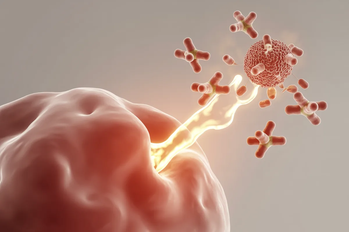
An allergic reaction is, at its core, an immune system reacting to something harmless as though it were a threat. The medical term is a hypersensitivity reaction: an exaggerated or inappropriate immune response to an antigen the patient has become sensitive to. Most of what we call “allergy” in clinical practice — the sneezing, the watery eyes, the hives, the wheal that rises on a skin test — runs through one specific immunologic pathway built around two players: the mast cell and the molecule it releases, histamine.
This guide is part of Empire Medical Training's Allergy Testing & Treatment resource center and is written for clinicians who want the mechanism, not just the vocabulary. Understanding it well is what lets you read a skin test confidently, explain antihistamines to a patient, and recognize when a reaction has crossed from nuisance into emergency. It is clinical education, not medical advice, and not a substitute for hands-on training.
What a hypersensitivity reaction is
As Dr. Sherry Wehner frames it in Empire's course, allergy is “a hypersensitivity reaction that occurs when a patient is exposed to an antigen that he or she is sensitive to.” The key word is sensitive. The antigen — a grass pollen, a cat dander protein, a peanut protein — is not inherently dangerous. The problem is the immune response to it. In a hypersensitive patient, the immune system has been primed to treat that antigen as a target, and it mounts a reaction out of proportion to any real threat.
This is not a rare quirk. Allergic disease affects more than 56 million Americans. Roughly 30% of U.S. adults have environmental allergies and at least 10% have a food allergy, and allergy ranks as the sixth leading cause of chronic disease in the country. Like most disease, it is multifactorial — the product of genetics, environment, and immunology together. The genetic component is striking: a child with one allergic parent is 30–50% more likely to develop allergies, and with two allergic parents, 60–80% more likely. What follows is the immunology piece — the cellular machinery that turns a genetic and environmental predisposition into a sneeze, a hive, or a wheal.
The type-I IgE-mediated pathway
The classic allergic reaction is a type-I, IgE-mediated hypersensitivity. It unfolds in two acts separated in time, which is exactly why a patient can tolerate a substance once and react to it later.
First exposure: sensitization
On the very first meaningful encounter with an antigen, the patient usually feels nothing. Behind the scenes, though, the immune system processes the antigen and B cells produce a specific class of antibody: IgE. That IgE does not float around doing damage. Instead it travels to mast cells in the tissues and basophils in the blood and binds to high-affinity receptors on their surface, coating them with antigen-specific antibody. The patient is now sensitized — armed and waiting — but still asymptomatic. Sensitization is the quiet first act of every type-I allergy.
Re-exposure: cross-linking and degranulation
The next time that antigen appears, it meets a mast cell already studded with matching IgE. The antigen bridges two adjacent IgE molecules — an event called cross-linking — and that bridging is the trigger. Cross-linking signals the mast cell to degranulate: it dumps the contents of its pre-formed granules into the surrounding tissue within minutes. Chief among those contents is histamine, released alongside other mediators including prostaglandins, tryptase, and heparin. This is the moment the patient finally feels the allergy — and because the granules are pre-made, the reaction is fast.
What histamine does in tissue
Histamine is the headline mediator because it produces, almost single-handedly, the signs and symptoms clinicians recognize as allergy. Once released into tissue it acts on local blood vessels, nerves, and smooth muscle:
- Vasodilation → redness. Histamine relaxes the small blood vessels, increasing blood flow to the area. Clinically this is the flare — the red halo around a reactive site, the flushed conjunctiva, the reddened skin.
- Increased vascular permeability → swelling. Histamine causes endothelial gapping — the cells lining the capillaries pull apart slightly — so fluid leaks out of the vessels into the tissue. That fluid is the wheal, the raised bump on skin testing; the same leak produces the nasal congestion, the puffy eyelids, and the soft-tissue swelling of an allergic reaction.
- Sensory nerve stimulation → itch. Histamine directly stimulates the sensory nerve endings that signal pruritus. This is the itch of hives, the itchy nose and eyes, the maddening itch of an insect reaction.
- Smooth-muscle contraction → bronchoconstriction. On airway smooth muscle, histamine and the other mediators can drive bronchoconstriction, a contributor to the airway component of an allergic reaction and part of why allergy and asthma are so closely linked.
Histamine does not act alone — prostaglandins, tryptase, and heparin contribute to the vascular leak and swelling as well — but if you remember one molecule, remember histamine. The familiar triad of itch, redness, and swelling is histamine writing its signature on the tissue.
Early-phase and late-phase response
The reaction described so far — cross-linking, degranulation, histamine — is the early-phase response. It begins within minutes of re-exposure and is dominated by the pre-formed mediators released from the granules. It is fast, and it is what a skin test captures in its fifteen-minute reading window.
Many patients then experience a late-phase response hours later. After the initial burst, the activated mast cells and recruited immune cells release newly synthesized mediators and chemical signals that draw in additional inflammatory cells, producing a second wave of swelling and congestion that can persist for hours. Clinically this is why a patient's symptoms can flare again in the evening after a morning exposure, and why allergic inflammation can feel stubborn rather than fleeting. The early phase is histamine's opening act; the late phase is the broader inflammatory response settling in.
Why the wheal-and-flare appears on skin testing
This single pathway is what makes allergy skin testing work. A skin prick test introduces a tiny amount of allergen into the superficial skin. In a sensitized patient, local mast cells are already coated with antigen-specific IgE; the introduced allergen cross-links that IgE, the mast cell degranulates, and histamine pours into that one small spot of skin. You then watch the type-I reaction play out in miniature:
- The wheal — the raised, pale swelling at the center — is histamine-driven vascular leak (endothelial gapping) in action.
- The flare — the red halo around it — is histamine-driven vasodilation.
This is also why testing is read against controls. A positive histamine control confirms the skin is capable of mounting a wheal, and a negative (glycerin) control establishes the patient's baseline; a result is read as positive when the wheal is at least 3 millimeters larger than the negative control. The mechanism even explains a common pitfall: in dermatographism, mast cells in the superficial dermis release histamine in response to friction or scratching rather than to any antigen, so the patient develops a wheal-and-flare wherever the skin is stroked — which is why the negative control can come up positive and why such patients are better served by blood testing. The exact technique, controls, panel selection, and interpretation are taught hands-on in Empire's course.
Why antihistamines work — and interfere with testing
Once you see histamine as the engine of the reaction, the entire logic of treatment falls into place. Antihistamines block the histamine receptors on the very tissues histamine acts upon — the blood vessels and the sensory nerves — so the signal arrives but the tissue can no longer respond to it. With those receptors occupied, vasodilation, vascular leak, and itch are all blunted, which is why an antihistamine relieves the runny nose, the watery itchy eyes, and the hives of an allergic reaction.
That same mechanism creates a practical problem the clinician must respect: antihistamines suppress the wheal-and-flare and will cause false-negative skin tests. A truly sensitized patient who is still taking an antihistamine can test negative simply because the skin cannot mount the visible reaction. For this reason patients are generally asked to stop antihistamines for several days before testing, and the positive control is checked first to confirm the test is even valid. (Other agents — certain antidepressants and topical steroids among them — can blunt the response too; which medications to hold and for how long is covered in the course.)
The four hypersensitivity types, briefly
For context, type-I is one of four hypersensitivity types in the classic Gell and Coombs classification. Most clinical allergy is type I, but the others matter at the edges of allergy practice:
- Type I — immediate, IgE-mediated. The pathway described throughout this page: mast cells, histamine, the wheal-and-flare. The mechanism behind environmental and IgE-mediated food allergy.
- Type II — antibody-mediated cytotoxic. Antibody binds to cells and marks them for destruction, resulting in tissue damage.
- Type III — immune-complex mediated. Antibody-antigen complexes deposit in tissue and provoke inflammation. This is a delayed, IgG-associated mechanism relevant to some delayed food reactions.
- Type IV — delayed, T-cell-mediated. Not antibody-driven at all; T cells produce the reaction over a day or more. This is the mechanism of contact dermatitis, which is why patch testing — read at 48 hours — is fundamentally different from the immediate prick test.
The distinction is more than academic. Knowing that contact dermatitis is type IV rather than type I tells you immediately why an antihistamine helps the hives but not the poison-ivy rash, and why the two are tested completely differently.
Safety, scope, and when to refer
The same pathway that produces a harmless wheal can, in its most extreme form, produce anaphylaxis — a massive, systemic mast-cell release affecting the airway, circulation, and skin at once. Anaphylaxis is a medical emergency, and first-line treatment is intramuscular epinephrine. Because skin testing deliberately provokes the type-I reaction, it must always be performed with emergency preparedness on hand — epinephrine available and staff trained to use it — even though serious reactions to testing are rare.
This is clinician education, not a tool for patient self-diagnosis, and certain patients belong with a board-certified allergist or immunologist rather than a general practice: those with a history of anaphylaxis, severe or uncontrolled asthma, or prior systemic reactions to testing. Testing and treating allergy responsibly requires proper training, the right clinical setting, and genuine emergency readiness — the focus of Empire's Allergy Test & Treatment training. The immunology on this page also connects to the broader functional medicine view of immune dysregulation and root-cause inflammation.
Master the science behind the wheal
Empire Medical Training's Allergy Test & Treatment training teaches the immunology, skin and blood testing, immunotherapy, and the in-office allergy lab — taught by Dr. Sherry Wehner, MD, who built allergy testing into her own practice. Understand the mechanism, then learn to apply it safely and profitably.
Explore the Allergy Training →