
Skin testing is the workhorse of allergy diagnosis in a general practice. When a patient presents with the classic picture — allergic rhinitis, chronic sinusitis, conjunctivitis, eczema, or suspected food or insect allergy — a well-run skin-prick test can confirm which allergens the patient is sensitized to in about fifteen minutes, in the office, at low cost. That speed and visibility are exactly why it remains the first-line tool for environmental allergy, and why it is one of the most accessible procedures a non-specialist clinician can add to practice.
This guide situates skin testing within the broader field of allergy testing and treatment and is written for clinicians who want an accurate, practical overview. It is clinical education, not medical advice, and nothing here should be read as a treatment recommendation, protocol, or substitute for hands-on training and current clinical guidance.
How allergy skin testing works
Skin testing is a direct readout of the allergic cascade. In a sensitized patient, allergen-specific IgE antibodies sit bound to receptors on mast cells in the skin. When the test introduces that allergen into the superficial skin, it cross-links the IgE, the mast cell degranulates, and it releases mediators — most importantly histamine. Histamine then produces the three findings clinicians recognize at the bedside: nerve stimulation causing itch, vasodilation causing the surrounding redness (the flare), and endothelial gapping that lets fluid leak out and raise the central wheal. Prostaglandins, tryptase, and heparin contribute to the swelling as well, but histamine is the headline mediator.
Because the test measures this physical response, it is essentially an in vivo assay: you are watching the patient's own mast cells answer the allergen in real time. That is its strength and its limitation. It is most sensitive for environmental allergens — pollens, dust mites, animal dander, molds — and is a reasonable, if less sensitive and specific, tool for IgE-mediated food allergens. It detects sensitization, not disease, which is why interpretation always loops back to the clinical history.
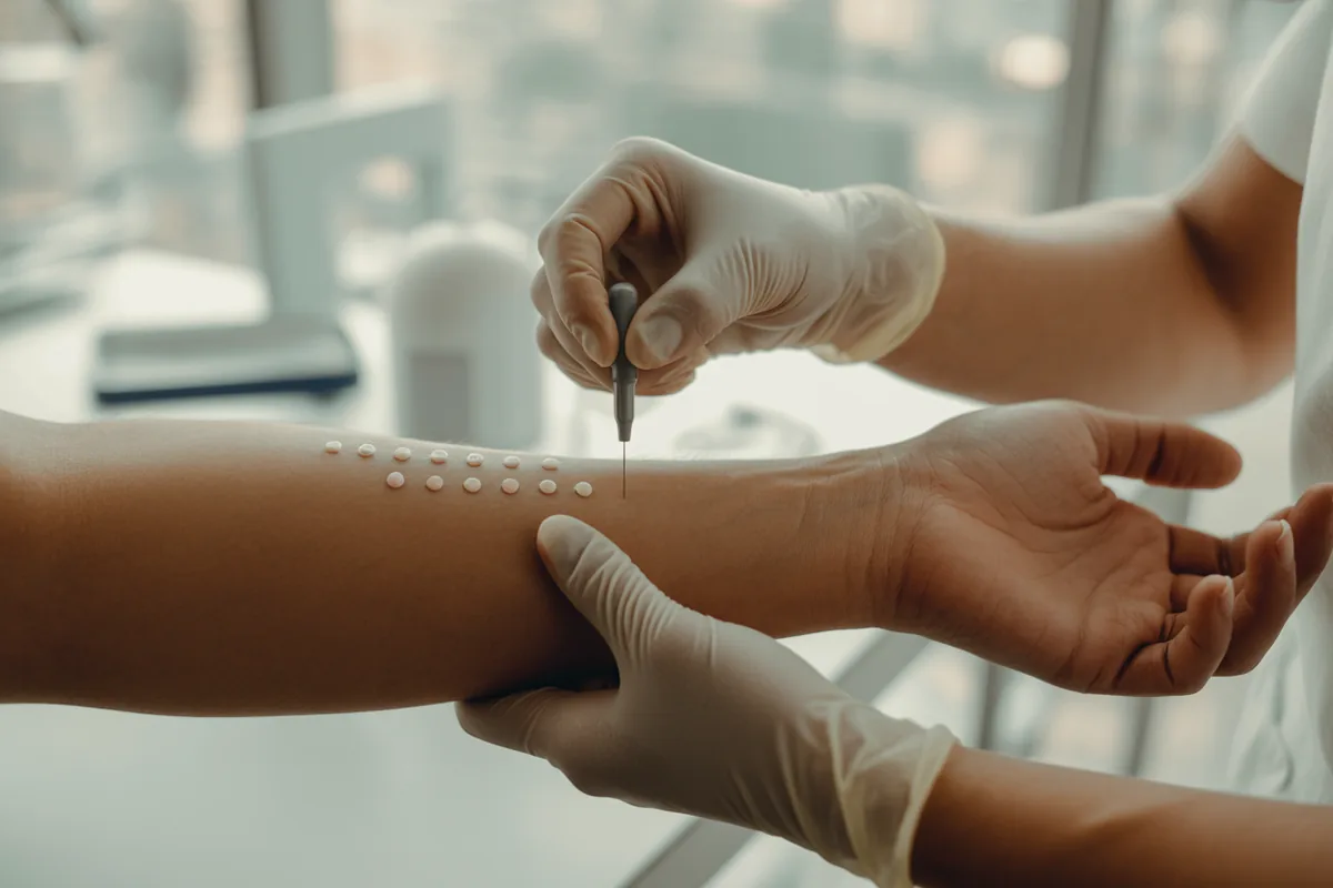
The skin-prick (percutaneous) test
The skin-prick test — sometimes called the percutaneous or puncture test — is the routine method. A small amount of each allergen extract is placed on the skin and a device pricks through it into the superficial epidermis, deep enough to deliver the allergen but not deep enough to draw blood. Testing is performed on the forearms and/or the back, the most reactive sites; the wrist is notably less reactive and is avoided.
Historically each allergen was dropped on the skin and pricked with a separate sterile lancet. Modern practice uses multi-headed plastic applicators that deliver many antigens at once in an even, well-spaced grid — faster, more consistent, and less prone to the bleeding that can cause false positives. A typical comprehensive panel tests dozens of environmental and food antigens in a single sitting; in Dr. Wehner's practice the standard panel runs to 76 antigens across both forearms and the back, always including controls. The whole panel is performed at once, because insurance generally reimburses skin-prick testing only once per year — a partial panel followed by a second visit will not be covered.
After application the test is left for about 15 minutes, then read. The patient may report itching at reactive sites, which is expected. The exact applicator technique, panel design, and site mapping are demonstrated step by step in Empire's hands-on course.
Controls: why a test is valid or not
No skin test can be interpreted without its controls. Every panel includes a positive control — histamine — and a negative control — the glycerin/saline diluent the extracts are suspended in.
- The positive (histamine) control must react. If it does not produce a wheal, the test is invalid — most often because the patient is still taking an antihistamine or another suppressing medication. You cannot trust a negative result when the positive control is flat.
- The negative control should be flat. If the saline/glycerin control raises a wheal, the patient likely has dermatographism — mast cells releasing histamine in response to mechanical friction rather than allergen — and every site will look falsely positive. Dermatographism affects roughly 4–5% of people and is a reason to switch to blood testing.
Only when the controls behave — positive reactive, negative flat — do the allergen results mean anything.
Reading the wheal-and-flare response
Results are read at about 15 minutes by measuring the largest diameter of the wheal. The flare (the surrounding redness) can be recorded, but immunotherapy and reimbursement decisions are anchored to the wheal. The practical threshold: a reaction counts as positive when the wheal is at least 3 millimeters larger than the negative control.
This 3-millimeter rule matters because many sites raise a small wheal that simply mirrors the negative control — those are negative, not positive, no matter how they look at a glance. A real positive is clearly larger than the negative control. In one of Dr. Wehner's own demonstrations, a patient with a mild dermatographic negative control still showed a clean, unambiguous mountain-cedar positive at roughly 1.5 cm because it stood well above the baseline reaction — a reminder that you read every result relative to that patient's own negative control, not against an absolute number.
Older plus-grading systems (1+ through 4+) exist but are not the basis for reimbursement and are no longer recommended; measured wheal diameter is the standard. The full reading and documentation workflow — what to chart, how the technician records each reagent, and how positives feed immunotherapy eligibility — is covered in the course.
What interferes with results
A skin test is only as good as the conditions it is run under. The dominant variable is medication:
- Antihistamines are the classic confounder and must usually be stopped about four to five days before testing. They blunt the histamine response and produce false negatives — including a non-reactive positive control. If a patient cannot or will not stop them, that points toward blood testing.
- Tricyclic antidepressants and topical steroids applied to the test area can also cause false negatives. Inhaled corticosteroids do not interfere, so a patient can stay on their nasal or inhaled steroid; short courses of systemic corticosteroids, however, can suppress the response.
- Beta blockers and ACE inhibitors deserve special mention. They do not necessarily distort the test, but beta blockers can blunt the response to epinephrine if a reaction occurs, and ACE inhibitors are associated with a more exaggerated allergic response — both are reasons to refer such patients to a board-certified allergist rather than test them in a general practice.
Other factors shift sensitivity too: the quality of the allergen extract, the body site used, and patient age — testing in patients over 50 tends to be less reactive. Technique errors matter as well: sites placed too close together can't be read accurately, induced bleeding can cause false positives, and insufficient skin penetration causes false negatives.
Prick vs intradermal testing
The intradermal test injects a small amount of allergen into the superficial dermis rather than pricking it into the surface. It is more sensitive but less specific, generating more false positives (and some false negatives), which is why in routine human allergy practice the skin-prick test is the primary tool and intradermal testing is used selectively — classically after a negative prick test for high-stakes allergens such as venoms or certain drugs. The table below summarizes the practical trade-offs.
| Feature | Skin-prick (percutaneous) | Intradermal |
|---|---|---|
| Technique | Allergen placed on skin, pricked into superficial epidermis | Allergen injected into the superficial dermis |
| Sensitivity | Good; lower than intradermal | Higher |
| Specificity | Higher — fewer false positives | Lower — more false positives |
| Routine use | First-line for environmental and food allergens | Selective — e.g., after a negative prick for venoms/some drugs |
| Throughput | Many antigens at once via multi-head applicator | One site at a time; slower |
| Risk profile | Very low; reactions rare | Higher allergen load delivered, so somewhat greater reaction risk |
A related method, patch testing, is a different test for a different mechanism: it screens for T-cell–mediated contact dermatitis (metals, cosmetics, preservatives) rather than IgE-mediated allergy, leaving allergen-loaded patches on the skin for 48 hours. It is covered as its own topic in specialized allergy testing.
Skin testing vs blood testing
The main alternative to skin testing is specific-IgE blood testing (commonly called RAST). Each has appropriate uses, and neither is a stand-alone diagnosis without clinical correlation. The advantages of skin testing are practical and real: it is fast, low-cost, performed in-office, and gives visible same-visit results. Blood testing comes into its own precisely where skin testing fails — when a patient cannot stop interfering antihistamines, has severe eczema or dermatographism that makes the skin unreadable, is a very young child, or has a history of a systemic reaction to skin testing. It can also screen a larger number of antigens at once.
The decision is rarely about which test is "better" in the abstract; it is about which test the patient in front of you can actually be tested with, and what the history suggests. For a full treatment of the serology option, see blood testing for allergies. To understand which allergens these tests are screening for in the first place, see types of allergens, and for the clinical picture that prompts testing, our overview of allergy symptoms.
Safety and emergency preparedness
Skin testing is very safe, but "very safe" is not "risk-free," and the distinction is the whole point of this section. Severe reactions are genuinely rare: in a frequently cited Mayo Clinic series of roughly 500,000 skin tests, only six patients had systemic reactions, none were severe, and all resolved within an hour. Most reactions are nothing more than local erythema and itching that settle with a little topical corticosteroid.
Even so, systemic reactions and anaphylaxis are possible, and testing must never be performed without emergency preparedness on hand. The first-line treatment for anaphylaxis is intramuscular epinephrine, and an EpiPen or equivalent auto-injector should be immediately accessible and familiar to staff before a single prick is placed. Equally important is patient selection: a history of anaphylaxis, severe or uncontrolled asthma, or medications such as beta blockers and ACE inhibitors that complicate emergency management are all reasons to refer to a board-certified allergist or immunologist rather than test in a general office. Severe eczematous dermatitis on the test site, dermatographism, pregnancy, and very young age are additional cautions. The specific emergency protocol, dosing, and staff readiness drills are taught in Empire's course.
Learn allergy skin testing the right way
Empire Medical Training's Allergy Test & Treatment Training teaches the skin-prick technique, panel design, reading and documentation, intradermal and patch testing, immunotherapy, and the in-office allergy lab — with a live skin-test demonstration by Dr. Sherry Wehner, MD. CME-accredited, with a practical, add-it-to-your-practice focus.
Explore the Allergy Training →