







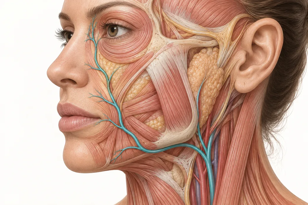
The most common failure in occlusion recognition is not ignorance of the signs. It is looking in the wrong place for them. An injector treats a nasolabial fold, something feels wrong, and they stare at the nasolabial fold. Meanwhile the finding that would have made the diagnosis is on the nasal ala, two centimeters away, in a vascular territory nobody is examining because nobody put a needle there.
Skin changes happen outside of where you injected. That is not an occasional curiosity; it is the expected behavior of an obstructed arterial supply, and an assessment confined to the puncture site is structurally incapable of finding it.
Three reasons the finding moves
1. The deposit is not at the entry
The first displacement is mechanical and it is the smallest of the three. Product sits at the needle tip, generally around half an inch beyond the entry point for most filler placement and considerably further with a cannula. Before any vascular physiology is involved, the material in question is already somewhere other than the mark on the skin.
2. Ischemia presents downstream
The second displacement is the important one. When a vessel is obstructed, the tissue that suffers is the tissue it was supplying — the territory distal to the point of obstruction, downstream in the direction of flow. The obstruction itself is invisible. The territory is what you can see.
This means the distance and direction between your needle and your findings are determined by the arterial anatomy, not by the geometry of your injection. A small-caliber terminal branch produces a small, well-demarcated patch nearby. A larger, more proximal vessel produces a wider territory further away. In some cases the material travels retrograde — against flow, under injection pressure — and then washes forward into a completely different distribution when pressure is released. That is the mechanism behind the most feared presentations, including ophthalmic involvement from midface and nasal injections, which we cover in our piece on whether injectables can cause blindness.
3. Inflammation and spasm track along the vessel
The third displacement runs the other way. Inflammation and vessel spasm can extend along the vessel proximally — back toward the source — so tenderness, induration and erythema may be appreciable along the vascular course, not just in the ischemic territory downstream.
The result is that a single event can produce findings in three separate locations: at the deposit, along the vessel, and in the distal territory. A clinician examining only one of the three will see a partial picture and may well see nothing.
The anastomotic complication
Facial vasculature is richly interconnected, and the anastomoses cut both ways for the injector.
In your favor: collateral supply is why many small occlusions cause limited damage, why some territories tolerate partial obstruction without visible change, and why outcomes vary so widely between patients with similar events.
Against you: anastomoses are also the pathway by which material reaches territories that have no obvious relationship to the injection site. The connection between the external and internal carotid systems through the angular and dorsal nasal vessels is the reason a filler placed in the midface can affect the ophthalmic circulation. Anastomotic crossover is why the scan has to be anatomical rather than local — you are following a network, not a line.
The detailed arterial mapping of the face is its own body of work and belongs alongside this piece rather than inside it. What matters for recognition is the operational consequence: before you inject, you should be able to name the vessels supplying the region, the direction they run, and the territory each one feeds.
Why some territories fail faster than others
Collateral supply is not evenly distributed, and the regions with the least of it are the ones where an occlusion converts to tissue loss quickest.
The nasal ala and tip are the standing example: the supply is functionally end-arterial in character, the skin is tight, and there is little redundancy to rescue the territory. The glabella behaves similarly — a comparatively isolated supply, tethered tissue and a short route to the ophthalmic circulation. In territories like these a modest obstruction produces a disproportionate result, and the window in which intervention changes the outcome is correspondingly short.
The practical consequence for assessment is a threshold, not a technique. The same equivocal finding warrants faster action on a nasal ala than on a well-collateralized cheek, because the cost of a cautious extra fifteen minutes is not the same in the two locations.
What to scan, by region
This is a scanning checklist for perfusion assessment, not an anatomy reference. For each region you treat, these are the additional areas that must be included in the look.
| Treated region | Also scan |
|---|---|
| Nasolabial fold, medial cheek | Nasal ala and sidewall, nasal tip, upper lip, infraorbital skin |
| Nose (dorsum, tip, ala) | Entire nose including columella and both alae, glabella, medial canthal skin — and ask about vision |
| Glabella, medial brow | Forehead, medial brow, upper eyelid skin — and ask about vision |
| Lips | Full vermilion, both sides, philtrum, perioral skin, chin |
| Infraorbital, tear trough | Lower eyelid skin, medial cheek, nasal sidewall — and ask about vision |
| Temple | Temporal scalp, lateral forehead, lateral brow, upper eyelid |
| Chin, jawline | Chin, lower lip, submental skin |
Three rules travel with the table.
Always include the contralateral match in the scan. Color and temperature findings are comparisons. Scanning only the treated side removes the reference that makes a subtle change visible.
Ask about vision whenever you have treated anywhere in the midface, nose, glabella or periorbital region. This is a question, not an observation, and it is easy to forget in a room where everything else is visual. Any visual symptom is an immediate escalation with no watch-and-wait.
Scan under consistent, neutral lighting. A subtle dusky change that is obvious under one light source disappears under another.
The conceptual switch: territories, not points
The mental shift this asks for is from thinking about injection sites to thinking about supply regions.
An injection site is a point on a face chart. A territory is a volume of tissue with an inflow, an outflow, a set of collaterals and a set of neighbors it exchanges with. Once you are thinking in territories, several things that seem puzzling become obvious.
Why does the nasal tip go dusky after a nasolabial injection? Because those structures share a supply and the obstruction was proximal to both.
Why is the finding larger than the treated area? Because the territory is larger than the treated area; the size of the ischemic zone reflects the vessel involved, not the volume injected.
Why does one patient show a dramatic reticular pattern and another show a small pale patch from apparently similar events? Because collateral supply and the caliber of the obstructed vessel differ, and the territory reflects both.
Why is the entry point almost never where the action is? Because the entry point is a surface landmark with no vascular meaning at all.
This is also the anatomical reasoning behind treatment. When reversal is indicated, the target is the tissue containing the product and the affected territory — you flood the territory, oriented to where the tip was rather than to the puncture. Hyaluronidase crosses tissue planes and the vessel wall, so intraluminal placement is not required and should not be attempted under duress. Our discussion of dissolving filler covers the enzyme; territory thinking is what tells you how much tissue you are trying to saturate.
This reflects Michelle Langston's clinical practice as taught in Empire Medical Training's hands-on curriculum. Technique is learned under supervision; this article is educational and is not a substitute for training.
How this changes the appointment
Pre-injection. Before the first pass, name the territory out loud, or at least deliberately: these are the vessels here, this is where they run, this is what they supply, and this is the area I will scan if anything is off. Ten seconds of anatomical orientation converts a later scan from a search into a check.
During and immediately after. Scan the territory, not the site — before the patient sits up and again before they leave.
At the same-day contact. Ask about the territory rather than the treatment area. Not "how is the cheek?" but "does the skin around your nose or lip look blotchy or different in color, and is any part of it cool?" Patients report what they are asked about.
In the documentation. Record what was scanned, not just what was treated. "Territory scanned including nasal ala and upper lip, capillary refill and temperature equal to contralateral" is a clinically useful record. "No blanching at injection site" is not.
Territory thinking is anatomy applied at the chairside, and it is learned the way anatomy is learned — by seeing it. Empire's Anatomical Based Aesthetics Training and Special Anatomical Cadaver Aesthetics Training put clinicians in front of the structures directly, and the Master Eye & Nose Injection Training addresses the regions where territory reasoning matters most.
Related guides in this cluster
Part of Vascular Occlusion: Recognition and Response.
 Clinical GuideThe Expected Filler Injection Response — Building the Baseline That Lets You Recognize IschemiaThe expected filler injection response — erythema, edema, ecchymosis — and how a trained normal baseline makes early ischemia recogniza
 Clinical GuideThe Golden Triangle — Why the Filler Deposit Site Isn't Where You Put the Needle InThe filler deposit site sits at the needle tip, not the entry point — about half an inch away, further with a cannula. Why that gap dec
 Clinical GuideImmediate, Early and Late — The Three Windows of Vascular Occlusion OnsetVascular occlusion onset is not one moment. Organize the picture into immediate, early and late windows — detection runs days past the
Train with Empire
This guide is clinical education. The technique behind it is taught hands-on, on live patients, with faculty beside you.
Explore Complete Dermal Filler Training →Disclaimer
This article reflects the clinical opinions and experience of Michelle Langston, APRN, MSN, FNP-BC, an independent faculty member contributing to Empire Medical Training's curriculum. The views expressed are the author's own and do not necessarily represent those of Empire Medical Training.
It is professional education, not medical advice, and is no substitute for hands-on training or independent clinical judgment. Licensed clinicians remain responsible for their own patient selection, technique and outcomes, for verifying current product labelling, and for practising within their scope and applicable law. Empire Medical Training accepts no liability for reliance on this content.
 Claim Your Prize!