







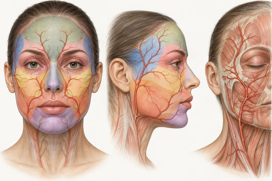
Vascular occlusion is not a rare event that happens to careless injectors. It is an uncommon event that happens to careful ones, and the difference between a complication that resolves and a complication that scars is almost never the moment of injection. It is the twenty minutes after it.
What separates injectors who manage occlusion well is not that they recognise it faster in some intuitive sense. It is that they built the comparison in advance. They know what this face looked like before the needle went in, they know which territory the vessel they are near actually supplies, and they have a sequence they run rather than a feeling they follow.
This page is the map of that discipline. Every section below opens into a deeper guide.
Recognition begins before the injection
You cannot detect a deviation from normal if you never established normal. The single most common failure in occlusion recognition is not missing a dramatic sign — it is having nothing to compare a subtle one against.
Three findings are expected after filler and should not alarm anyone: transient blanching from volume, pinpoint bleeding, and immediate mild oedema. Knowing precisely how each behaves, and over what timescale, is what lets you notice the one that is behaving wrongly. That is the subject of the expected filler injection response, and it is genuinely the first thing to learn in this cluster.
There is a second piece of pre-injection reasoning that changes everything downstream. The place the product ends up is not the place the needle entered. Entry point, deposit site and vascular territory are three different locations, and the gap between them is where most diagnostic confusion lives. The golden triangle explains why an injector who reasons from the entry point will look in the wrong place, and why a cannula changes that geometry without removing the problem.
The three windows of onset
Occlusion does not have one presentation. It has three, separated by time, and each is detected differently.
Immediate — during the pass itself. Pain out of proportion, blanching that does not refill, a patient who reacts differently than they did thirty seconds ago.
Early — the hours afterwards, when the appointment is over and the patient has gone home. This is the window that depends entirely on what you told them to watch for.
Late — days rather than hours, which is the window most injectors do not believe in until they see one.
Each window has a different detection method and a different realistic ceiling on how fast you can act. The three windows of vascular occlusion onset works through all three, including the one presentation that does not fit any window at all.
The perfusion exam
When something looks wrong, the first action is not to reach for hyaluronidase. It is to stop injecting and run a sequence.
A structured perfusion exam is five steps: scan the whole vascular territory rather than the injection site, ask about pain and its character, check capillary refill, compare temperature against the contralateral side, and re-examine over a defined interval rather than once. Running it the same way every time is what makes the findings interpretable — a capillary refill you have checked a thousand times tells you something; one you check for the first time under stress does not.
The five-step perfusion exam is the sequence itself, in order, with what each step is actually testing.
Read the territory, not the point
This is the conceptual switch that does the most work in this entire cluster.
Arterial supply is territorial. When a vessel is compromised, the finding appears wherever that vessel supplies — which may be centimetres from where you injected, may cross the midline, and may appear in a region you never touched, because the facial vasculature is a communicating network rather than a set of independent pipes.
An injector scanning the injection site will miss it. An injector scanning the territory will find it. Read the vascular territory, not the injection point covers what to scan by region and how it changes the shape of the appointment.
Two case-derived guides extend the same principle. The asymmetry tell shows how the distribution of a finding — and crucially, which areas are spared — can tell you whether you are looking at an occlusion or something else. Read the sequence, not the snapshot makes the case that a single photograph of a mottled lip is close to uninterpretable, and that morphologic progression over hours is the diagnostic signal.
Compression is not the same thing, and you can have both
Volume placed into a confined space can compress a vessel from outside it. That is a different mechanism from intravascular embolus, and the distinction matters because the two behave differently over time.
It also matters because the honest answer is usually that you cannot fully separate them at the chairside, and that treating for the more serious possibility is the correct posture. Compression occlusion versus vascular occlusion covers what the distinction does and does not change about your response, and why volume per pass is the control variable you actually hold.
Hyaluronidase: the mechanism, and the boundary
Hyaluronidase is the reason hyaluronic acid fillers are as forgiving as they are. Understanding why it works changes how you use it.
The enzyme recognises a substrate. It degrades hyaluronic acid, and the degree of cross-linking in a given product changes how readily it does so — which is why "the dose" is a less coherent idea than it sounds, and why treating the territory rather than the visible spot follows from the mechanism rather than from a protocol. What hyaluronidase actually does covers substrate, cross-linking, and passive movement across the vessel wall.
The boundary matters just as much. There is no substrate in a non-HA filler. Calcium hydroxylapatite, poly-L-lactic acid, polymethylmethacrylate and permanent products are not touched by the enzyme. That does not mean you withhold it — but it does mean your escalation threshold moves earlier, because the thing that usually buys you time is not available. What hyaluronidase cannot do is the guide that most changes practice for injectors who use more than one product family.
Specific hyaluronidase dosing, reconstitution and re-dosing intervals are taught hands-on in the workshop rather than published here. The reasoning is public; the protocol belongs in a room where it can be supervised.
Triage, transfer and the end of the emergency
Most occlusion management begins with a phone call, and the call arrives before any diagnosis is possible. Sorting it into routine, urgent or emergency — before you know what it is — is a separate skill from diagnosing it. Triaging the post-filler phone call sets out the three tiers and the questions that assign them.
If a case needs to leave your clinic, how it leaves determines what happens next. The rules are unglamorous and absolute: never send a patient to an emergency department alone with instructions, keep treating while you wait, speak clinician-to-clinician rather than letting the patient carry the history, and do not let anyone leave until perfusion is documented. What a competent emergency transfer looks like is the protocol.
Finally, the question nobody asks until they are in it: when is this actually over? Perfusion returning is the first endpoint, not the last. Vascular occlusion recovery and monitoring covers the daily recheck, what can still go wrong in week one, the decision to retreat an unresolved case, and why the hyperbaric oxygen pathway is something you build before you need it rather than search for at 9pm.
How to use this cluster
If you are starting from nothing, read in this order: the expected response, the golden triangle, the onset windows, then the perfusion exam. Those four give you recognition. The hyaluronidase pair gives you the intervention and its limits. Triage, transfer and recovery give you the parts that happen after the decision.
None of it replaces supervised practice. Recognition is a trained observation, and it trains fastest against real tissue with someone standing next to you.
Every guide in this cluster
 Clinical GuideThe Expected Filler Injection Response — Building the Baseline That Lets You Recognize IschemiaThe expected filler injection response — erythema, edema, ecchymosis — and how a trained normal baseline makes early ischemia recogniza
 Clinical GuideThe Golden Triangle — Why the Filler Deposit Site Isn't Where You Put the Needle InThe filler deposit site sits at the needle tip, not the entry point — about half an inch away, further with a cannula. Why that gap dec
 Clinical GuideImmediate, Early and Late — The Three Windows of Vascular Occlusion OnsetVascular occlusion onset is not one moment. Organize the picture into immediate, early and late windows — detection runs days past the
 Clinical GuideThe Five-Step Perfusion Exam — Reading the Signs of Vascular Occlusion in SequenceThe signs of vascular occlusion are only reliable inside a fixed sequence: scan, pain trajectory, capillary refill, temperature, vascul
 Clinical GuideCompression Occlusion vs Vascular Occlusion — and Why You Can Have BothCompression occlusion and intravascular occlusion are different mechanisms that coexist. Why volume per pass is a perfusion decision, n
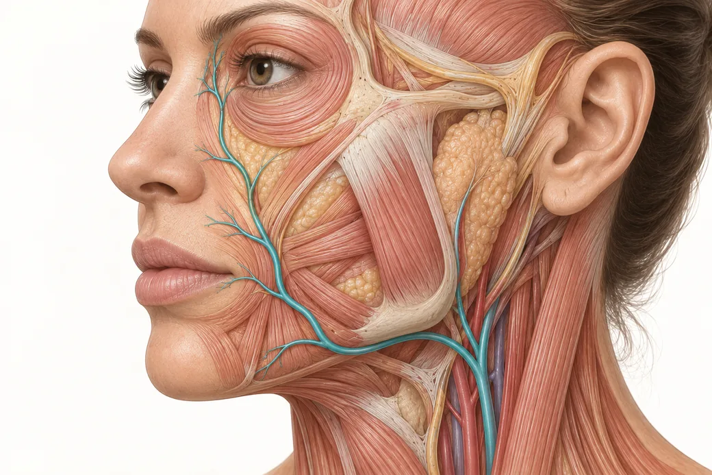 Clinical GuideRead the Vascular Territory, Not the Injection Point — Why Findings Appear Away From the DepositOcclusion findings appear in the vascular territory, not at the puncture. Scan downstream, know why inflammation tracks proximally, and
 Clinical GuideWhen Is the Emergency Actually Over? Vascular Occlusion Recovery and the Re-Injection WindowVascular occlusion recovery is measured in weeks, not minutes. Daily rechecks, retreating an unresolved case, blistering, and the re-in
 Clinical GuideRoutine, Urgent or Emergency: Triaging the Post-Filler Phone CallPost-filler phone call triage in three tiers — routine, urgent, emergency. Michelle Langston's framework for deciding before you have a
 Clinical GuideWhat Hyaluronidase Actually Does: Enzyme Mechanism for InjectorsHyaluronidase mechanism of action for injectors: cross-link cleavage, tissue permeability, passive vessel-wall transit, and why you flo
 Clinical GuideWhat Hyaluronidase Cannot Do: Non-HA Filler ComplicationsHyaluronidase does not reverse CaHA or PLLA. Why Michelle Langston still floods the territory, and why a biostimulator complication is
 Clinical GuideThe Handoff: What a Competent Emergency Transfer Looks LikeFiller complication emergency transfer, done properly: never send the patient alone, keep treating while EMS comes, and what the ED phy
 Clinical GuideThe Asymmetry Tell: When Vascular Occlusion Asymmetry Matches Your Injection PatternVascular occlusion asymmetry is diagnostic data. Why sparing of untreated tissue argues against generalised trauma and for a perfusion
 Clinical GuideRead the Sequence, Not the Snapshot: Occlusion Morphologic Progression in the Upper LipOcclusion morphologic progression in one site — dusky spot, blanching, mottling, spread, reticular change, dusky discolouration — and w
Train with Empire
This guide is clinical education. The technique behind it is taught hands-on, on live patients, with faculty beside you.
Explore Complete Dermal Filler Training → Claim Your Prize!