







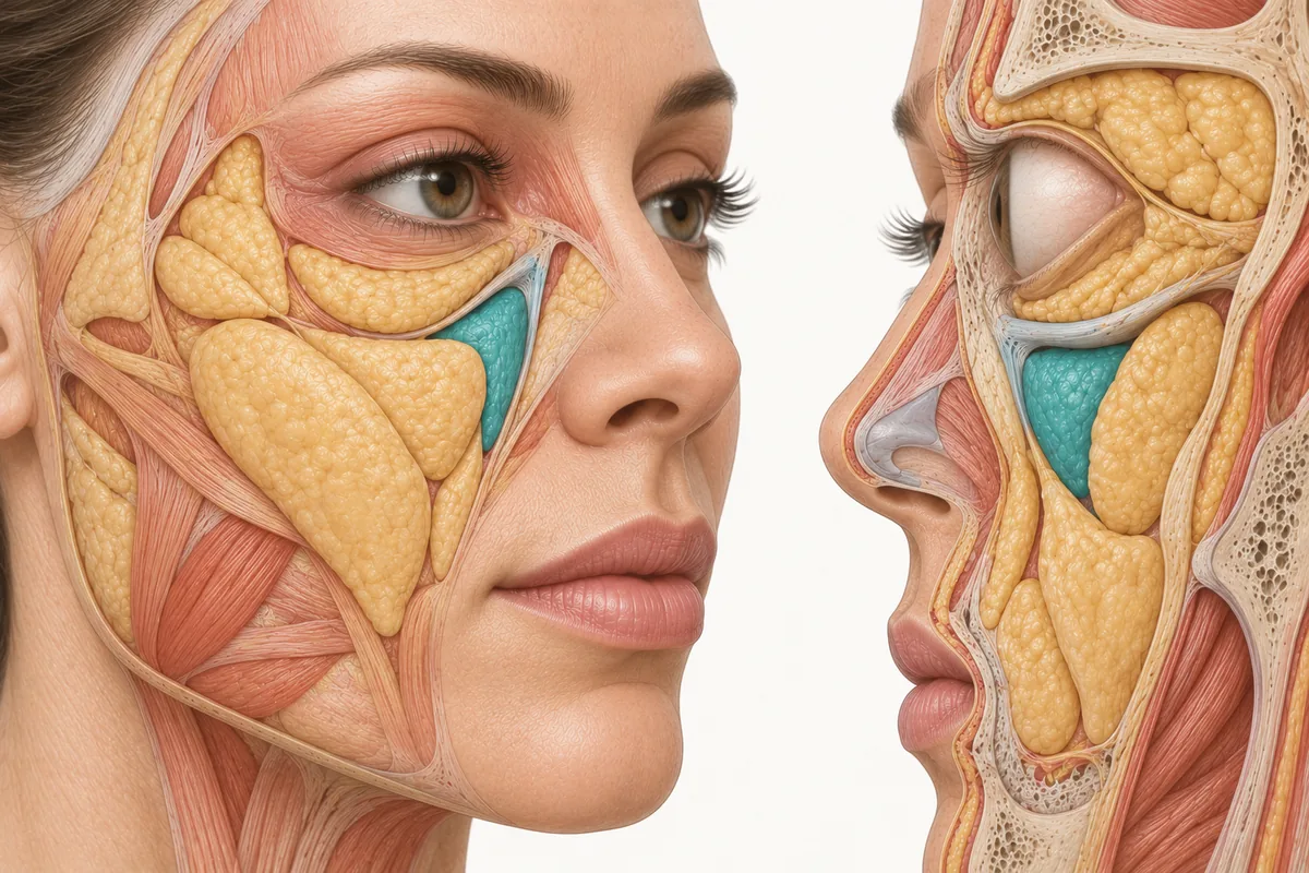
Ask why clinicians reach for PRF for the tear trough rather than PRP and the usual answer is about growth factors. That answer is incomplete and it is not the operative one. The operative reason is mechanical: PRF is the more viscous of the two materials, it clots rather than disperses, and in a region defined by thin skin and unforgiving tissue planes those two physical properties change what a deposit does after you have made it.
This article is about the material and the instrument. It is deliberately not a technique article, and the reason for that is stated plainly further down.
PRF vs Tear Trough Filler: the Material Question Comes First
The infraorbital region punishes the wrong material more than almost anywhere on the face. The skin is the thinnest on the body, the subcutaneous tissue is minimal, and anything that disperses unevenly, sits superficially, or holds water will be visible through the overlying skin rather than hidden by it.
A liquid material distributes along the path of least resistance. That is an advantage when you want broad coverage across a whole cheek or forehead. In a shallow, sharply-defined hollow bordered by structures you do not want to treat, it is a liability — the deposit goes where the tissue lets it, not where you intended.
A material that gels in situ behaves differently. It resists dispersion, occupies the space it was placed into, and holds its platelet and leukocyte cargo locally rather than allowing it to wash into surrounding planes. That is the property Tatiana Sarmiento is selecting for when she teaches PRF as her choice for this region in Empire Medical Training's curriculum: she describes the material as more viscous than PRP, identifies that viscosity as what makes it usable for targeted support, and names cannula delivery as her preferred approach for it.
These figures reflect Tatiana Sarmiento's clinical practice as taught in Empire Medical Training's hands-on curriculum. Technique is learned under supervision; this article is educational and is not a substitute for training.
PRF Viscosity: What “Thicker” Actually Means in the Syringe
Viscosity in PRF is not a fixed number. It is a moving one, and that is the part clinicians underestimate.
PRF contains no anticoagulant. Fibrin polymerisation begins as soon as blood contacts the tube wall and continues while the material sits in your syringe. The product is therefore more viscous than PRP at the moment you draw it up, and progressively more viscous with every minute that passes. The material you deliver in your last syringe is not the material you delivered in your first.
Three consequences follow directly.
Plunger force rises during the case. Resistance you interpret as technique is often just polymerisation advancing. Rising resistance is a signal to stop and reassess, never a signal to push harder — in thin-skinned anatomy, a forced deposit is an uncontrolled deposit.
Lumen resistance becomes the limiting factor. Pushing a thickening material through a narrow, long lumen requires high force, and high force is exactly what removes your ability to modulate delivery. The instrument has to accommodate the material.
The window governs the plan. Whatever the treatment involves, it has to be completable inside the working window of a material that is actively setting. That constrains how much can sensibly be attempted in one sitting, and it is a scheduling fact as much as a clinical one.
Why Use a Cannula for Tear Trough PRF
The rationale for a blunt cannula over a sharp needle in the infraorbital region is not specific to PRF — it is the general argument for cannula use in vascular-dense, thin-skinned anatomy, and it rests on two independent properties.
The blunt tip. A blunt-tipped cannula preferentially displaces mobile structures rather than penetrating them. It does not make vascular events impossible, and no instrument does, but it changes the probability. This is the same reasoning that governs instrument selection anywhere the consequence of an intravascular deposit is severe — a logic every injector should already have internalised from the vascular risk literature on facial injection.
A single entry point. One entry, from which a region can be reached, means fewer punctures through thin skin and fewer opportunities for bruising in tissue where bruising is conspicuous and slow to resolve.
To those general arguments PRF adds a third, material-specific one. A cannula's lumen accommodates a viscous material at lower plunger force than a fine needle of comparable length, which preserves the injector's ability to modulate delivery rather than fighting the syringe. When the material is actively thickening, that margin is not a luxury.
That is the whole instrument rationale. Which cannula, entering where, and travelling in which plane are questions with real answers, and none of them belong in an article.
What the PRF Under-Eye Evidence Actually Shows
The honest summary is that this is a promising, actively-researched application with real limitations, not a settled one.
A 2025 systematic review of 14 studies of injectable platelet concentrates for periorbital rejuvenation found PRF associated with improvements in skin texture, wrinkles and crepiness, with a favourable safety profile — mild, transient adverse effects and high patient satisfaction. One conclusion from that review belongs in every consultation: PRF improvements often diminished by six months (Sollitto et al., Journal of Cosmetic Dermatology, 2025).
Ready to put this into practice?
Explore Empire's hands-on, CME-accredited Functional & Regenerative Medicine courses — live patients, expert faculty, and ongoing mentorship.
Work on adjacent periocular applications is consistent with that shape. A retrospective series of 24 eyes in 12 patients treated with injectable PRF for superior sulcus hollowing across three sessions reported a significant reduction in sulcus depth at three months, mild regression by six months, 75% patient satisfaction and no recorded adverse effects (Arslan et al., Journal of Cosmetic Dermatology, 2026). Improvement, then partial regression — the same curve.
A parallel line of development combines denatured albumin with liquid PRF to produce a more structured injectable gel for periocular use, positioned as an autologous alternative for patients seeking a conservative approach or with prior hyaluronic acid complications, with progressive improvement reported over repeated treatments (Rahbari et al., Facial Plastic Surgery, 2026). Worth knowing this exists; it is a distinct preparation with its own protocol, not interchangeable with liquid PRF.
Two practical readings for the consultation. First, a series is the unit of treatment, not a single visit. Tatiana Sarmiento teaches a three-session approach for this region, and the published series that report meaningful change are also multi-session. Second, maintenance should be discussed before the first treatment, not after the result fades. If the published pattern is improvement at three months with regression by six, a patient told to expect a durable single-visit correction has been mis-sold.
PRF Tear Trough Before and After: Why the Photos Mislead
Tatiana Sarmiento makes an observation about photography in this region that is worth taking seriously, because it cuts both ways.
Her point is that a PRP facial result photographs as a subtle skin-quality change — tighter-looking pores, better tone, a refreshed appearance that a patient may struggle to see in a side-by-side. A PRF result in the tear trough photographs as something else entirely: a visible change in contour and in apparent colour, the kind of before-and-after a patient reads instantly as dramatic.
That is a real difference and it is not an artefact. Filling a shadow changes what the camera records twice over — the contour itself, and the darkness that the contour was casting. An area that looked pigmented can look lighter without any pigment having changed.
Which is exactly where the honesty problem begins.
A photograph that looks like structural correction invites the patient to hear a structural promise. It is not one. A platelet concentrate is a resorbing autologous material, not a filler; it does not restore lost structural volume, reposition tissue or correct laxity, and if the clinical job is genuinely volume replacement then the honest answer is a different category of product. Understanding where facial volume is actually lost — and the real distinction between stimulating tissue and replacing it — is what keeps that conversation straight.
A photograph also invites the patient to expect that result to persist. The published evidence says it frequently does not persist at six months. If your consent conversation and your gallery tell different stories, the gallery will win, and the dissatisfied patient is the one who believed it.
What this article is not
This is a product and instrument article. It contains no entry points, no tissue planes, no depths, no volumes and no injection sequence, and that omission is deliberate.
The infraorbital region is high-risk anatomy. It sits in a vascular territory where an intravascular deposit carries consequences that are not recoverable with an apology, and the margin between an acceptable plane and an unacceptable one is measured in millimetres of tissue you cannot see. Technique in this region is learned in person, on supervised cases, with an instructor's hands available — not from a written description, however carefully written.
If you intend to treat this area, get trained in it specifically. Empire Medical Training's Master Eye and Nose Injection Training covers injection of the periorbital and nasal regions, and Platelet Rich Plasma Training covers preparation and application of autologous platelet concentrates. The material reasoning above will make you a better-informed trainee. It will not make you a trained one.
Frequently Asked Questions
Why is PRF preferred over PRP in the tear trough?
Primarily for mechanical reasons. PRF is more viscous and gels in situ rather than dispersing, so a deposit resists spreading into surrounding planes in a region where uneven distribution is visible through very thin skin. Its sustained release profile is a secondary argument; the material's clot behaviour is the operative one.
Does PRF in the tear trough work as a filler substitute?
No. PRF is a resorbing autologous matrix, not a volumising product. It does not restore lost structural volume, reposition tissue or correct laxity. Photographs can look like structural correction because filling a shadow changes both contour and apparent darkness, but the underlying mechanism and the durability are different.
How long do periorbital PRF results last?
Current systematic review evidence reports that PRF improvements in this region often diminish by six months, and a published series on adjacent periocular hollowing showed significant improvement at three months with mild regression by six. Treatment is realistically a series with planned maintenance, and that should be said before the first appointment.
Why use a cannula rather than a needle for PRF here?
Three reasons combine. A blunt tip preferentially displaces rather than penetrates mobile structures in vascular-dense anatomy; a single entry point means fewer punctures through conspicuous thin skin; and a cannula's lumen passes a thickening viscous material at lower plunger force, preserving control as the material continues to polymerise.
Disclaimer
This article reflects the clinical opinions and experience of Tatiana Sarmiento, faculty, Empire Medical Training, an independent faculty member contributing to Empire Medical Training's curriculum. The views expressed are the author's own and do not necessarily represent those of Empire Medical Training.
It is professional education, not medical advice, and is no substitute for hands-on training or independent clinical judgment. Licensed clinicians remain responsible for their own patient selection, technique and outcomes, for verifying current product labelling, and for practising within their scope and applicable law. Empire Medical Training accepts no liability for reliance on this content.



 Claim Your Prize!