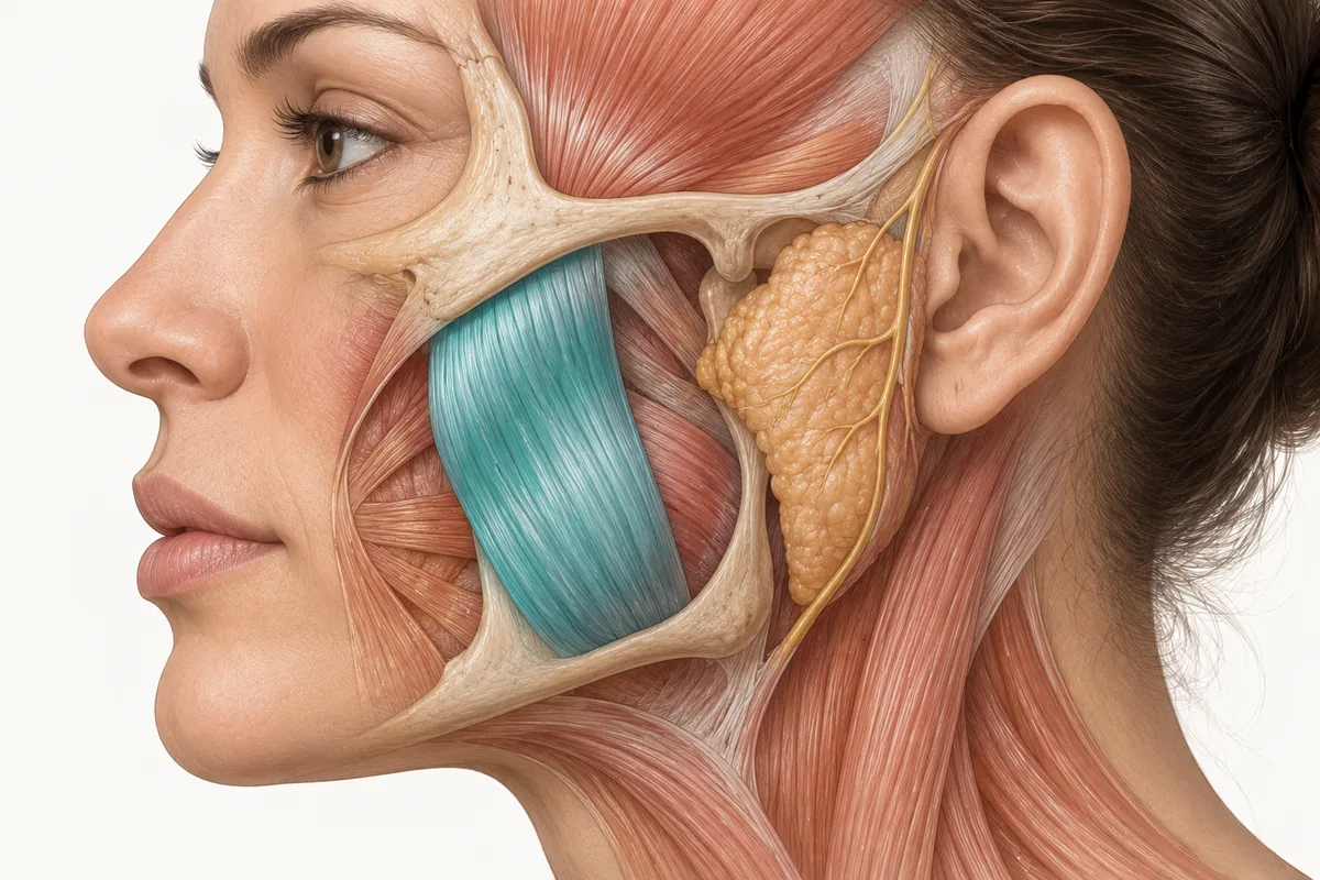
A masseter hypertrophy differential diagnosis is the thing standing between you and the most avoidable failure in lower-face neurotoxin: treating a muscle that is not the reason the jaw looks wide. If the width is skeletal, the toxin does nothing to it. If the fullness is a parotid gland, the toxin does nothing to it. And in a small number of cases the swelling you were asked to slim is a lesion that needed a diagnosis rather than an injection.
My honest position is that this is not a common trap. Most patients arrive with a reasonable understanding of masseter hypertrophy, and we rarely see them with a frankly wrong diagnosis. But the consequence of missing it is disproportionate to how often it happens — an unnecessary treatment, a patient who paid for nothing, and occasionally a delayed diagnosis. The screening costs you ninety seconds.
The ninety-second screen: does it contract?
Everything starts with the clench-release-palpate examination. Place your fingers over the masseter body bilaterally and have the patient clench, release, clench, release while you palpate and watch the muscle function.
A masseter changes. It hardens, it bulges, it recruits under your fingers and it softens when they let go. Nothing else in that anatomical region does. A parotid gland does not contract. A bony prominence does not contract. A lipoma does not contract.
So the first question is not "is this masseter hypertrophy?" but "does the thing making this face wide change consistency when the patient bites down?" If the answer is no, or if the swelling persists unchanged through the whole sequence, you are looking at something else, and neurotoxin is not the answer to it.
The second question is where the fullness sits. Masseter bulk sits over the ramus and angle of the mandible, bounded by the palpable anterior and posterior borders of the muscle. Parotid fullness sits more posteriorly and superiorly, overlying and behind the posterior masseter and extending toward the ear and the retromandibular region. Bony width is at the angle itself, hard, immobile and identical at rest and on clench.
These figures reflect Dr. Croley's clinical practice as taught in Empire Medical Training's hands-on curriculum. Technique is learned under supervision; this article is educational and is not a substitute for training.
The published differential
Masseter hypertrophy is usually an asymptomatic enlargement of one or both masseters, and in most cases no aetiological factor is identified, so it is regarded as idiopathic. It also leads to a prominent mandibular angle that patients find aesthetically unacceptable — which is how it arrives in an aesthetic clinic rather than a surgical one.
The differential reported in the oral and maxillofacial literature for enlargement in this region includes parotitis, parotid tumour, lipoma, benign and malignant muscle tumours, vascular tumours, and benign and malignant mandibular tumours, along with salivary gland disease more broadly and masseteric myopathy. Accurate diagnosis is explicitly noted to be more difficult in unilateral cases, and bilateral masseteric hypertrophy is itself listed as something to consider in the differential of unilateral or bilateral swelling of the parotid or lateral mandibular area — the confusion runs in both directions (Shetty et al., Case Reports in Dentistry, 2012; see also the masseteric hypertrophy case literature indexed in PMC).
Two entries on that list are common enough to matter to an aesthetic practice.
Parotid enlargement
If there is a genuinely enlarged parotid gland, treating the masseter will not slim the lower face. That is a mechanical certainty, not a probability.
The parotid overlies the posterior portion of the masseter, so gland enlargement and muscle hypertrophy produce fullness in overlapping but not identical locations. The clinically useful distinction is still the clench: the gland does not change with contraction, and it tends to displace the fullness posteriorly and inferiorly toward the angle of the jaw and the earlobe rather than over the muscle belly.
The benign cause most likely to walk into an aesthetic clinic is sialadenosis, also called sialosis — a non-inflammatory, non-neoplastic, usually bilateral salivary gland disorder presenting as chronic, persistent, painless, largely asymptomatic parotid hypertrophy. It is consistently associated with an underlying systemic condition: alcoholism, diabetes, bulimia and other eating disorders, endocrine disorders, malnutrition and pregnancy are the classically reported associations, with the proposed unifying mechanism being an autonomic peripheral neuropathy of the glandular nerve supply leading to acinar cell engorgement (University of Iowa Head and Neck Protocols, Sialosis or Sialadenosis of the Salivary Glands).
That association list is why this matters beyond aesthetics. Bilateral painless parotid enlargement in a young patient requesting facial slimming can be the presenting sign of an eating disorder. A referral is the correct response; a syringe is not.
The other parotid entries — parotitis, and benign or malignant parotid neoplasms — are not aesthetic diagnoses at all. Unilateral, firm, nodular, painful, rapidly enlarging, fixed to deeper tissue, or accompanied by any facial nerve weakness: that patient needs imaging and a head and neck opinion, not neurotoxin.
Bone
This is the one that most often produces a disappointed patient rather than a dangerous one.
The mandibular skeleton governs the shape of the lower third of the face, and the contour of that region is determined by both the shape of the mandible and the bulk of the masseter. In the mandibular contouring literature — largely written about square-face correction in Asian populations, where the problem has been studied most systematically — the skeletal contribution is frequently described as the more prominent element, with masseteric hypertrophy present but not dominant. Surgical planning in that literature separates the two causes explicitly: mandibular angle ostectomy is chosen when the prominent angle is caused by a reduced gonial angle seen on profile view, while outer-cortex ostectomy is chosen when the prominence is caused by lateral protrusion of the angle. Formal assessment for these procedures evaluates ramus height, gonial angle, mandibular width, divergence and masseter thickness together, because they are separate contributors to the same silhouette (mandibular contouring and square-face categorisation literature, Journal of Plastic, Reconstructive & Aesthetic Surgery and related sources).
Ready to put this into practice?
Explore Empire's hands-on, CME-accredited Aesthetic & Injectable Training courses — live patients, expert faculty, and ongoing mentorship.
You are not planning an osteotomy. But the logic transfers directly: on a patient whose bigonial width is largely bone, chemodenervating a modest masseter will produce a modest result, and the more square the bony angle, the smaller the proportion of the width you can influence. The clinical signs are a hard, sharply defined angle palpable through a relaxed masseter, lateral flare visible on the submental or oblique view, a low gonial angle on profile, and a masseter that does not feel especially bulky on maximal clench.
The right move here is not to refuse treatment. It is to quantify the expectation before you inject: some of this width is muscle and we can reduce that portion; some of it is the shape of your jawbone and neurotoxin does not change bone.
The less common entries
Lipoma, benign and malignant muscle and vascular tumours, and mandibular tumours all belong on the list even though you may go years without seeing one. So does unilateral mandibular condylar hyperplasia, which has been reported presenting primarily as parotid swelling with ipsilateral masticator muscle and parotid hypertrophy — a reminder that the region's structures enlarge together and that unilateral findings deserve more suspicion than bilateral ones (Springer, BMC Musculoskeletal Disorders, 2025).
The red flags that end the aesthetic consultation
Stop and refer rather than treat when you find any of:
- Unilateral enlargement without a clear history of unilateral chewing or clenching
- Rapid growth, or a swelling the patient reports as new and changing
- Pain, tenderness or overlying skin change
- A firm, fixed or nodular mass, or one that does not soften between clenches
- Any facial nerve weakness or new asymmetry of expression
- Trismus or progressive restriction of mouth opening
- Systemic features — weight change, xerostomia, dry eyes, lymphadenopathy, or a history consistent with the sialadenosis associations above
None of these are reasons for embarrassment. They are reasons for a referral letter.
What this changes at the chairside
Add three lines to your masseter consultation and you have covered almost all of it. Palpate through a clench-release cycle and ask yourself whether the fullness contracts. Palpate the bony angle through the relaxed muscle and decide how much of the width is skeleton. Ask whether the enlargement is bilateral and long-standing, or unilateral and new.
Then say out loud, before you inject, which portion of the width you expect to change. The patient who is told "most of this is muscle and we can reduce it" and the patient who is told "some of this is the shape of your jaw and it will not change" are both satisfiable. The patient who is told nothing and given an injection is the one who comes back unhappy at month six — and by then you have spent both their money and your credibility.
The regional anatomy underlying all of this is worked through in Empire's Anatomical Based Aesthetics Training and, in cadaveric detail, in Special Anatomical Cadaver Aesthetics Training. Where the masseter genuinely is the culprit, our material on botox for jaw clenching and management of bruxism covers the therapeutic indication.
About the author
Dr. Chris Croley — Chief Medical Officer, Empire Medical Training. The clinical approach described here is what he teaches in Empire's hands-on neurotoxin curriculum.
Frequently Asked Questions
How do you distinguish parotid enlargement from masseter hypertrophy?
Palpate through a clench-release sequence. The masseter hardens and bulges with contraction; the parotid does not change. Parotid fullness also sits more posteriorly and superiorly, toward the ear and retromandibular region, rather than over the muscle belly across the ramus and angle.
What causes bilateral painless parotid enlargement?
Sialadenosis is the classic non-inflammatory, non-neoplastic cause, and it is consistently associated with an underlying systemic condition — alcoholism, diabetes, bulimia and other eating disorders, endocrine disorders, malnutrition and pregnancy. Bilateral painless parotid swelling in a young slimming candidate warrants a referral rather than an injection.
Can botox slim a jaw that is wide because of bone?
No. Neurotoxin reduces muscle bulk and has no effect on the mandibular skeleton. In a patient whose bigonial width is substantially skeletal, treating the masseter produces a proportionally smaller result, which is why the muscular and bony contributions should be separated and explained before treatment.
What are the red flags in a patient asking for masseter slimming?
Unilateral enlargement without a unilateral chewing history, rapid growth, pain, overlying skin change, a firm fixed or nodular mass, any facial nerve weakness, trismus, or systemic features. Any of these should prompt imaging and a head and neck opinion rather than an aesthetic treatment.
Is masseter hypertrophy itself a diagnosis of exclusion?
Largely, yes. In most cases no aetiological factor is identified and the condition is regarded as idiopathic, and the published differential includes parotid disease, lipoma, muscle and vascular tumours and mandibular tumours. Diagnosis is noted to be considerably harder in unilateral presentations.
Disclaimer
This article reflects the clinical opinions and experience of Dr. Chris Croley, Chief Medical Officer, Empire Medical Training, an independent faculty member contributing to Empire Medical Training's curriculum. The views expressed are the author's own and do not necessarily represent those of Empire Medical Training.
It is professional education, not medical advice, and is no substitute for hands-on training or independent clinical judgment. Licensed clinicians remain responsible for their own patient selection, technique and outcomes, for verifying current product labelling, and for practising within their scope and applicable law. Empire Medical Training accepts no liability for reliance on this content.