







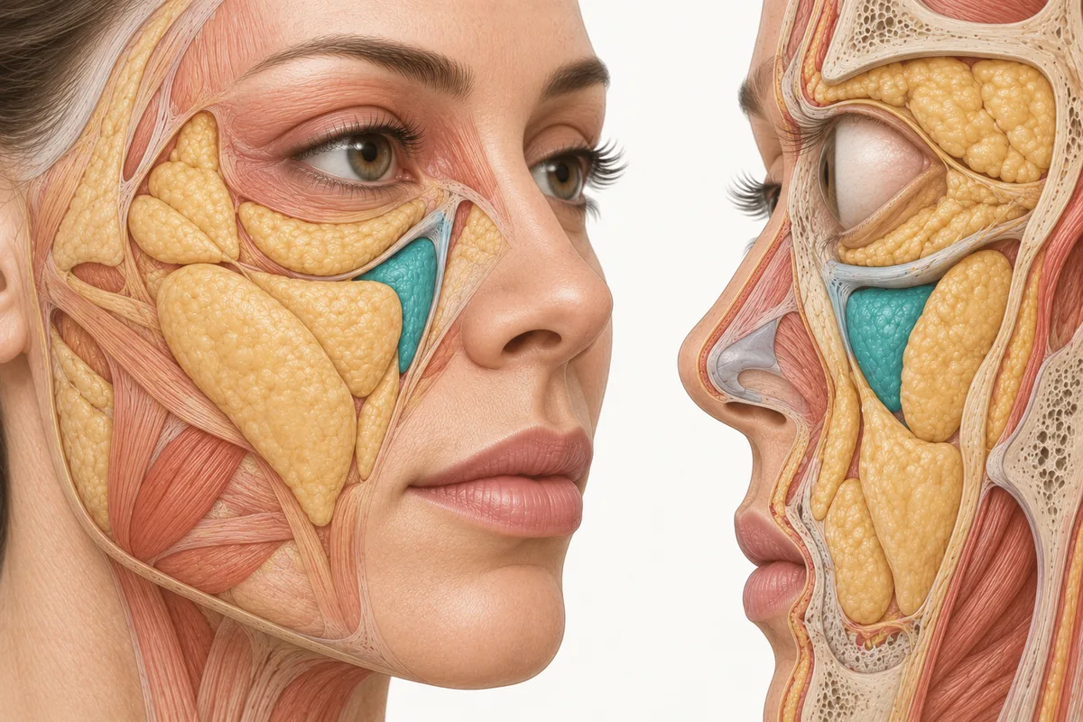
A patient sits down and says: I look tired. I hate my under-eyes. It is one of the most common opening lines in aesthetic practice, and it is a complaint, not a diagnosis. A useful tear trough differential diagnosis starts from the assumption that the patient has correctly identified where they see the problem and has told you nothing at all about where it comes from.
The worked example I teach is the one that surprises injectors most. Patient points at the infraorbital region. You assess, and what you find is loss of volume and projection across the zygomatic and medial cheek region. You support the midface — and the tear trough looks better, without anything having been placed under the eye at all. The complaint resolved because the driver was addressed. That is the entire argument for assessment over product selection, in one case.
Here is the differential that gets you there.
What Causes Tear Troughs: Nine Drivers Behind “I Look Tired”
1. A true infraorbital hollow. A genuine volume deficit in the infraorbital region, bounded below by a ligamentous attachment that does not move. This exists, it is the thing the patient thinks they have, and it is the minority of presentations.
2. Midface deficiency. Loss of deep medial cheek volume and malar projection lengthens the lid–cheek junction and creates a shadow at the transition. The under-eye looks hollow because the cheek stopped holding the light. This is the case above, and in my practice it is the single most common true driver of the complaint.
3. Skeletal change at the orbital rim and maxilla. The bony orbital aperture enlarges with age, with recession concentrated superomedially and inferolaterally while the central rim stays comparatively stable (Kahn DM, Shaw RB. Aesthetic Surgery Journal. 2008;28(3):258–264). The maxilla retrudes in parallel — the maxillary angle decreasing by around 10 degrees between young and older cohorts (reviewed in Mendelson B, Wong CH. Aesthetic Plastic Surgery. 2012;36(4):753–760). A patient with a receded rim and a retruded maxilla has a periorbital appearance that is a platform problem. Adding product superficially over a deficient platform tends to produce fullness without improvement.
4. Negative vector morphology. Some patients have this constitutionally rather than acquiring it: in profile, the anterior cornea sits ahead of the malar eminence rather than behind it. That morphology makes the infraorbital region structurally prone to hollowing and shadow, and it is a known predictor of a poor result from volume placed under the eye. Assess it in profile at the first visit, every time.
5. Pseudoherniation of orbital fat. Here the finding above the groove is a convexity, not a hollow — the septum has weakened and orbital fat is projecting forward. The trough looks deeper because the tissue above it is bulging. Filling the trough in this patient produces two convexities and a fuller, heavier lower lid.
6. Malar mounds and festoons. Fluid and lymphatic congestion overlying the malar region. They vary with time of day, salt, sleep and alcohol; they compress and refill. They are not a volume deficit, they respond poorly to volume, and volume placed nearby can make them worse. This is a decline-or-refer finding more often than it is a treatment target.
7. Periorbital hyperpigmentation. Pigment, not shadow. Constitutional, post-inflammatory, or allergic. No amount of structural support changes pigment, and this is one of the most common reasons an anatomically sound infraorbital plan disappoints a patient.
8. Skin quality at the lower lid. Crepiness, fine etched lines and laxity in the thinnest skin on the face. A surface-layer problem needing a surface-layer answer — and one that can coexist with any of the above.
9. Everything that is not the under-eye at all. Brow descent, upper lid hooding, and upper-face heaviness all read as "tired" to patients who then point at their lower lids. So does chronic allergic rhinitis, poor sleep, thyroid disease and dehydration. Ask.
Most patients have two or three of these simultaneously. The job of assessment is to rank them.
Dark Circles, Eye Bags or Sunken Eyes? The Tests That Separate Them
None of this requires equipment. All of it takes about ninety seconds.
Change the light. Assess the patient under overhead lighting and then with light coming from the front at eye level. A finding that softens or disappears when frontal light removes the shadow is a contour problem. A darkness that persists under flat frontal light is pigment. This single manoeuvre resolves the most consequential fork in the differential, and it is the one injectors skip most often.
Lie them supine. Have the patient lie flat and look again. Tissue that redistributes and fills the hollow when gravity is removed is telling you the problem involves descent and support. A hollow that is unchanged supine is a genuine contents deficit.
Lift the midface manually. Place a finger on the malar region and elevate gently, then ask the patient to look in the mirror. If the under-eye improves visibly when you support the cheek, you have your answer and your patient has watched you find it. This is also the most persuasive consultation tool I know, because it demonstrates the plan before it is proposed.
Stretch the skin. Gentle lateral traction on the lower lid skin. Pigment that persists on stretch is dermal or deeper. Darkness that vanishes is vascular or shadow.
Compress and watch it refill. For a suspected malar mound or festoon, apply light pressure and release. Fluid compresses and returns. Volume does not behave that way.
Assess the vector in profile. Look at the patient from the side. The relationship of the anterior cornea to the malar eminence takes two seconds to read and changes the risk profile of everything you might do afterwards.
Pinch and release. Standard laxity assessment, but do it at the lid–cheek junction specifically, where the skin is thinnest and where recoil tells you the most about the envelope.
Ask about the day. Findings that are worse in the morning and better by afternoon are fluid. Findings that are constant are structural.
Ready to put this into practice?
Explore Empire's hands-on, CME-accredited Aesthetic & Injectable Training courses — live patients, expert faculty, and ongoing mentorship.
How to Fix Tear Troughs: Rank the Drivers Before You Treat
Once you have the list, rank it: what is the dominant driver, what is contributing, and what is present but not causing the complaint.
Then say it out loud, in plain language, before you propose anything. Something close to: what you are seeing as a hollow under your eye is mostly a shadow, and the shadow is there because the cheek underneath has lost support. If I support the cheek, the shadow softens. Some of the darkness is pigment, and that will not change with any of this — that needs a different treatment, and I want to be honest that it may not fully resolve.
Two things happen when you talk this way. The patient consents to a plan that does not inject the area they pointed at, which is otherwise a very hard sell. And you have set an expectation about the component that will not improve, before rather than after treatment — which is the difference between a satisfied patient and a disappointed one at the two-week review.
When Tear Trough Filler Goes Wrong: Why the Under-Eye Is Unforgiving
Two reasons, and both argue for a conservative default in this region.
The periorbital area is a high-consequence vascular territory. Depth decisions here are risk decisions as well as aesthetic ones — the reason vision loss after facial injection is a periorbital and glabellar subject rather than a general one.
And the tissue is unforgiving of product that is misplaced or over-placed. The skin is the thinnest on the face, the lymphatic drainage is idiosyncratic, and product that sits too superficially or in excess is visible, persistent and difficult to disguise. Correction is possible — dissolving hyaluronic acid filler is a real option — but it is a correction, and the patient experiences it as one.
If ever there were a region where the safest, most natural result is the one the anatomy can support, it is this one. Go conservative, review at two weeks, and add if the anatomy asks for it.
This reflects Melissa Pulcini-Buttine's clinical practice as taught in Empire Medical Training's hands-on curriculum. Technique is learned under supervision; this article is educational and is not a substitute for training.
Tear Trough Assessment: What Changes on Monday
Stop accepting "tear trough" as the presenting diagnosis. It is the patient's word for a region, not for a pathology.
Run the light test on every under-eye consultation. It costs nothing and it is the highest-yield discriminator you have.
Do the manual midface lift in front of a mirror, every time. It converts your assessment into something the patient can see, and it makes a midface-first plan explicable.
And when the differential comes back dominated by pigment, fluid or a negative vector, say so and adjust the plan rather than proceeding with the one the patient came in expecting.
The anatomy behind all of this — the orbital rim, the lid–cheek junction, the midface compartments and the vessels running through them — is taught in tissue in Empire's Anatomical Based Aesthetics Training, and the periorbital region specifically in Master Eye & Nose Injection Training. Full-face assessment, which is where most of these complaints are actually solved, is covered in Complete Facial Aesthetic Training.
About the author. Melissa Pulcini-Buttine, PA, has practised as a physician assistant for two decades and has taught anatomy and physiology for approximately fourteen years. She is a faculty member at Empire Medical Training and the founder of an aesthetics practice in Greenwich, Connecticut.
Frequently Asked Questions
Why do some under-eye complaints improve after treating the cheek?
Because the hollow is often a shadow cast at a lengthened lid–cheek junction rather than a true infraorbital deficit. Restoring medial cheek and malar support shortens that transition and removes the shadow. The complaint resolves because the anatomical driver was addressed, not because the region the patient pointed at was filled.
How do I tell shadow from pigment under the eye?
Change the light. Assess under overhead lighting, then with frontal light at eye level. Contour-based darkness softens or disappears when frontal light removes the shadow; pigment persists. A gentle lateral stretch of the lid skin confirms it — pigment that remains on stretch is dermal or deeper.
What is a negative vector and why does it matter under the eye?
In profile, a negative vector describes an anterior cornea that projects ahead of the malar eminence rather than behind it. That morphology predisposes to infraorbital hollowing and shadow, and it predicts a poorer response to volume placed under the eye. It should be assessed in profile at the first consultation.
Are malar mounds and festoons treated with filler?
Generally no. They are fluid and lymphatic findings rather than volume deficits — they vary through the day, compress under pressure and refill. Volume placed in or near them frequently worsens their appearance. They are more often a reason to decline or refer than a treatment target for an injector.
What should I tell a patient whose under-eye darkness is mostly pigment?
Tell them before treatment, not after. Explain that pigment is a separate problem from contour, that structural support will not change it, and that it needs a different class of treatment with a realistic and sometimes partial outcome. Setting that expectation at the consultation is what prevents a technically good result from being experienced as a failure.
Disclaimer
This article reflects the clinical opinions and experience of Melissa Pulcini-Buttine, PA, an independent faculty member contributing to Empire Medical Training's curriculum. The views expressed are the author's own and do not necessarily represent those of Empire Medical Training.
It is professional education, not medical advice, and is no substitute for hands-on training or independent clinical judgment. Licensed clinicians remain responsible for their own patient selection, technique and outcomes, for verifying current product labelling, and for practising within their scope and applicable law. Empire Medical Training accepts no liability for reliance on this content.



 Claim Your Prize!