







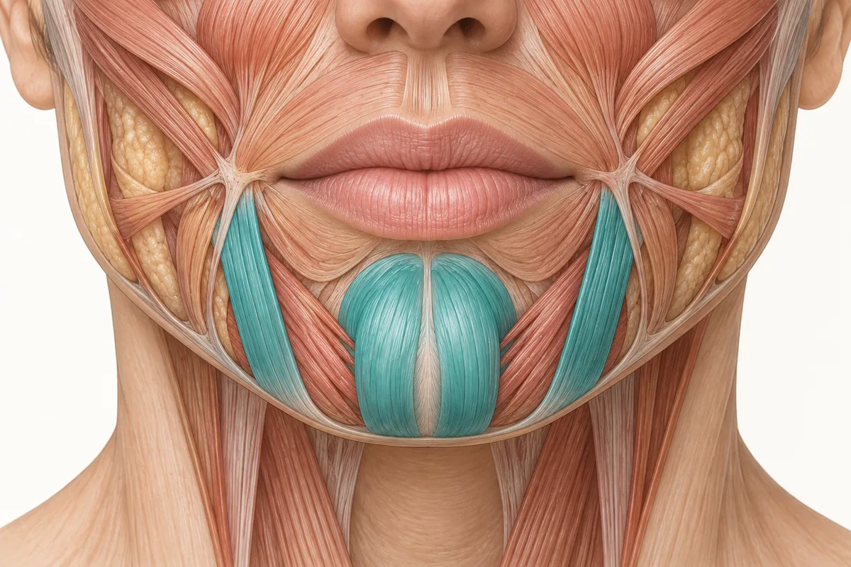
Search "facial danger zones" and you will find dozens of diagrams with red patches on a face. Injectors learn them, screenshot them, and carry them around as a mental overlay. I want to argue that the term should go, and that what replaces it — high-risk and low-risk — is not a softening of the language but a more accurate description of what is actually under the skin.
This is not pedantry. Terminology shapes cognition. The words you use to describe a risk determine what you notice, what you check, and what you stop checking. "Danger zone" teaches an injector three things that are not true, and the errors it produces are predictable.
Danger Zone Failure One: It Implies a Safe Zone
This is the big one. Every binary creates its complement. If a region is a danger zone, then by implication everywhere else is a safe zone — and that inference is wrong.
In anatomy, there is no 0% risk area. There is no region of the face that contains no vessels, no nerves, and no possibility of a poor outcome. There are regions where the vascular anatomy is more forgiving and regions where it is less forgiving, and that is a gradient, not a boundary.
The clinical failure mode this produces is specific and recognisable: an injector who is meticulous in the glabella and casual in the lower cheek. Slower, smaller aliquots, careful aspiration and deliberate planes in the "red" area — then speed, volume and autopilot everywhere else. The risk did not become zero when the needle crossed the edge of the diagram. The injector's attention did.
I have seen the same pattern in how people talk about complications afterward: "but it wasn't a danger zone." That sentence is a delay. It is a reason not to take a patient's report seriously, built entirely out of a word.
Danger Zone Failure Two: It Puts Risk in a Map, Not in Anatomy
A zone is a location. Anatomy is a structure.
When you teach a trainee that a patch of face is dangerous, you have given them a coordinate and nothing else. They cannot reason from it. They cannot extend it to a region you did not cover. They cannot adapt it to a patient whose anatomy differs from the diagram — which is every patient, since facial arterial anatomy is one of the most variable systems in the body.
When you teach that a region is high-risk because of what is underneath it, you have given them a mechanism. Mechanisms travel. A trainee who understands that the glabella is high-risk because it is a small territory hosting superficial branches of the ophthalmic artery which anastomose downward into the facial system can work out, unprompted, that the nasal dorsum is also high-risk — because it shares that plumbing. The zone diagram cannot do that.
This is the difference between memorising injection points and understanding why you are injecting where you are injecting. The zone map is the injection-point mentality applied to safety.
Danger Zone Failure Three: It Does Not Scale With the Variables
Risk in aesthetic injecting is not a property of a location alone. It is a product of at least five things:
- What vessel is likely underneath, and how variable its course is in that region
- What depth you are working at, and what occupies that layer there
- What you are injecting — rheology, particle behaviour, volume per aliquot
- How you are delivering it — needle or cannula, pressure, speed, static or moving tip
- What that territory connects to, which determines how far a problem can travel
A zone label collapses all five into one binary. The same square centimetre of face can be genuinely low-risk with a cannula in a supraperiosteal plane and genuinely high-risk with a sharp needle in a subdermal plane delivering a fast bolus. A red patch on a diagram cannot express that, so it teaches injectors to think about where and not about how.
High-risk and low-risk survive that complexity because they are relative terms. They invite the follow-up question — high risk relative to what, and modifiable by what? — that a zone label shuts down.
What Changes When High-Risk Replaces Facial Danger Zone
The vocabulary change is small. The behavioural change is not.
You stop switching attention on and off. Every region gets a survey. The survey is deeper and slower in high-risk territory, but it happens everywhere, because everywhere carries some risk.
You start giving reasons. "This is a high-risk area" is an incomplete sentence in a way that "this is a danger zone" is not. It prompts you to finish it: high-risk because the vessel here is shallow, because this territory connects to the ophthalmic circulation, because the anatomy here is unusually variable. Trainees can then be tested on the reason rather than the location.
You describe risk honestly to patients. "There are no zero-risk areas of the face; some carry more risk than others and here is why this one does" is a defensible, accurate consent conversation. "This is a danger zone" is alarming without being informative, and "this is a safe area" is a claim you cannot support.
You make your own limits explicit. If you are not yet confident with the vessels of a high-risk region and with your practice's occlusion response, the honest response is to be very cautious there or not to work there yet. That is a competency judgement, and it is much easier to make about a graded risk than about a red patch.
The three regions I teach as high-risk, and why
Three regions carry the highest risk in the anatomy I teach: the nose, the glabella and the midface. Note what they have in common — it is one thing, and it is not their position on a diagram.
Ready to put this into practice?
Explore Empire's hands-on, CME-accredited Aesthetic & Injectable Training courses — live patients, expert faculty, and ongoing mentorship.
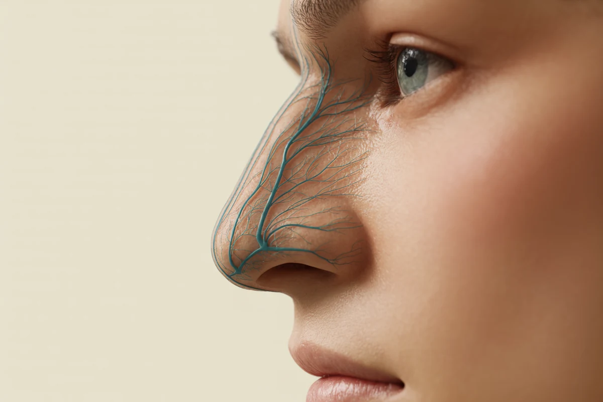
The nose. Supplied by the lateral nasal and dorsal nasal arteries, which anastomose with the angular artery below and the ophthalmic system above. It sits on the junction between the external and internal carotid territories. In the largest published review of filler-associated vision loss, the nose was the most common injection site at 40.6% of cases (Doyon et al., Aesthetic Surgery Journal, 2024;44(10):1091–1104).
The glabella. A small territory with a low margin for error, hosting the supratrochlear and supraorbital arteries — both branches of the ophthalmic artery, both running superficially through much of their course in this region — and communicating downward with the angular artery at the medial canthus.
The midface. Carries the angular segment of the facial artery toward the medial canthus, becoming progressively more superficial as it ascends. Published ultrasound work on the angular artery in the midface concludes plainly that there is no guaranteed safe location in this region (Cotofana et al., Aesthetic Surgery Journal, 2021;41(7):805–813).
That is the list. The reason each one is on it is anatomical: proximity to, or direct connection with, the arterial junctions that lead to the ophthalmic circulation. Learn the reason and the list stops being something you memorise.
What "low-risk" is actually claiming
It is worth being precise about the other half of the pair, because a lazy reading of "low-risk" reproduces exactly the problem we are trying to solve.
Low-risk means: the vascular anatomy in this territory is comparatively forgiving, the vessels are comparatively deep or comparatively distant from critical anastomoses, and an error here has a smaller ceiling of consequence. It does not mean no vessels. It does not mean no possibility of occlusion, nodule, infection or asymmetry. It does not mean stop looking.
It is a statement about the ceiling of the worst outcome, not about the probability of any outcome. That distinction is the whole point, and it is the part that "safe zone" destroys.
How to Explain Facial Injection Risk to a Patient
Patients ask about danger zones because that is the language online. You do not have to adopt it to answer well.
What works, in my experience, is naming the gradient honestly and then naming the controls: no area of the face is risk-free; some areas carry more risk than others because of the blood vessels beneath them; here is which category the area we are treating falls into and why; here is what I do differently because of it; and here is what I want you to call me about afterwards, including anything you notice in an area I did not treat.
That last clause matters more than the rest. Because arteries in the face are connected, a problem can present somewhere you never touched — which is precisely the thing a zone diagram trains a patient, and an injector, not to expect. For the separate question patients often raise about neurotoxin and vision, which has a different mechanism entirely, there is a dedicated article on whether botulinum toxin can cause blindness.
Teaching the Reason, Not the Facial Region
Empire Medical Training's anatomy curriculum is built around the reason rather than the map: what is underneath, in which layer, connected to what. Anatomical Based Aesthetics Training and Special Anatomical Cadaver Aesthetics Training work that reasoning in tissue, and Complete Facial Aesthetic Training applies it across the regions injectors treat most.
Change the words and the thinking follows. Risk in this work is a gradient with an anatomical explanation at every point on it — and an injector who carries that model checks more, assumes less, and recognises a problem faster than one carrying a picture with red patches on it.
Melissa Pulcini-Buttine, PA — physician assistant of two decades; professor of anatomy and physiology for ~14 years; faculty member, Empire Medical Training; founder of an aesthetics practice in Greenwich, Connecticut.
Frequently Asked Questions
What are facial danger zones?
"Facial danger zones" is the common term for facial regions where injection carries a higher risk of serious vascular complication. The term is widely used but poorly constructed: it implies the existence of safe zones, which do not exist anatomically. High-risk and low-risk describe the same anatomy more accurately, because facial risk is a gradient rather than a boundary.
Which areas of the face are highest risk for injectors?
Three regions carry the highest risk: the nose, the glabella and the midface. They share one property — each sits on or connects directly into the arterial junctions between the facial and ophthalmic circulations. Risk in these areas comes from the underlying vascular anatomy and its connections, not from position on a diagram.
Is any area of the face completely safe to inject?
No. There is no 0% risk area in facial anatomy. Every region contains vessels and nerves, and every injection carries some possibility of an adverse outcome. Low-risk describes a lower ceiling of worst-case consequence and comparatively more forgiving vascular anatomy — not the absence of risk, and not a reason to reduce vigilance.
Why does terminology matter for injection safety?
Because language determines attention. A binary "danger zone" label encourages injectors to concentrate in flagged regions and relax elsewhere, and it supplies a location without a mechanism, so it cannot be extended to unfamiliar territory. Risk language that names the anatomical reason produces reasoning an injector can apply to any region.
Disclaimer
This article reflects the clinical opinions and experience of Melissa Pulcini-Buttine, PA, an independent faculty member contributing to Empire Medical Training's curriculum. The views expressed are the author's own and do not necessarily represent those of Empire Medical Training.
It is professional education, not medical advice, and is no substitute for hands-on training or independent clinical judgment. Licensed clinicians remain responsible for their own patient selection, technique and outcomes, for verifying current product labelling, and for practising within their scope and applicable law. Empire Medical Training accepts no liability for reliance on this content.



 Claim Your Prize!