








I have taught anatomy and physiology for about fourteen years, and I can tell you exactly which lesson lands hardest. It is not a lecture. It is the moment an injector opens a second cadaver and finds an artery that does not match the first one.
Cadaver training for injectors is often described as an advanced add-on — something you do once you are established, a credential rather than a capability. I would put it differently. Dissection is the fastest way to convert anatomical knowledge into anatomical perception, and perception is what you actually use at the chairside. You do not consult a diagram while your hand is on a patient's face. You see, or you don't.
Here is what changes.
Cadaver Dissection Makes Anatomical Variability Real
Every anatomy text will tell you the facial artery is variable. You read it, you nod, you carry on picturing the diagram.
Then you dissect ten faces. One facial artery ends at the lip and never reaches the nose. One crosses the nasolabial fold twice. One is duplicated. One is so tortuous you lose it and find it again three centimetres higher. The left side of a face does not match the right side of the same face.
The published data say exactly this — one cadaveric series of 102 hemifaces documented eight different termination points and 35 distinct branching-pattern combinations, with the angular artery present in only about a third of specimens (Nguyen et al., Archives of Craniofacial Surgery, 2024;25(2):77–84). But reading "35 combinations" and finding your fourth unexpected arrangement in an afternoon are different events in a clinician's head.
After dissection, "the vessel is usually here" starts to sound like what it is: a probability statement. And an injector who hears probability builds margin, where an injector who hears fact does not.
Facial Layers Stop Being Bands of Color
A layered facial diagram shows five coloured stripes: skin, subcutaneous fat, SMAS, deep fat, periosteum. Clean, discrete, obvious.
Tissue does not present that way, and the difference is tactile. The retinacula cutis resist and then give. The areolar plane opens with almost no resistance at all — you feel the tissue separate rather than cut. An aponeurosis pushes back. Periosteum stops you.
That is what "know which layer you are in" actually means, and it is not learnable from an image, because the information is in the resistance and not in the appearance. Injectors who have dissected describe the same thing afterwards: they stop thinking about depth as a number on a needle and start thinking about it as a sequence of textures they are passing through.
It also makes regional variation concrete. The soft tissue envelope over the mid-cheek is generous; the envelope over the glabella is not. You can be told that. Passing a blade through both in the same session teaches it in a way that persists.
Injection Depth Becomes a Felt Distance, Not a Figure
Pooled anatomical data put the facial artery at roughly 6 mm from the skin at the mandibular border and 8–10 mm through the midface, while the terminal angular artery sits around 1 mm deep at the medial canthus. Those are useful numbers. They are also abstract until you have seen the difference with your own hands.
One millimetre at the medial canthus is a distance you can feel is nothing. Six millimetres at the mandible is a distance you can feel is real. After dissection, injectors calibrate — the numbers acquire a physical scale, and a needle depth stops being arithmetic.
The Facial Neighborhood Becomes Three-Dimensional
The last thing dissection does, and maybe the most important for safety, is convert a flat map into a neighbourhood.
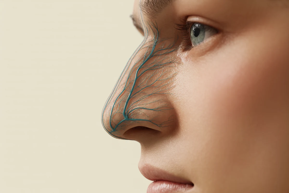
On a diagram, the angular artery and the dorsal nasal artery are two lines that meet. In tissue, at the medial canthus, they are two small vessels in a shallow, crowded space alongside the supratrochlear and supraorbital arteries, the medial canthal tendon, and the orbital rim — all within a couple of centimetres, all a millimetre or two from the surface.
Seeing that crowding is what makes the retrograde embolisation story stop being a paragraph you learned and start being a place you have been. Injectors come out of that dissection treating the medial canthus and the nasal base differently, permanently, without being told to.
What Cadaver Dissection Transfers to the Living Face
Five changes, and I watch for all of them in people who have done the work:
They survey before they plan. There is a pause before the first entry point that was not there before — a few seconds spent tracing the route under the skin.
They palpate. The facial artery pulse at the antegonial notch is patient-specific information available in seconds, and dissected injectors reach for it because they know exactly what they are feeling for.
They examine outside the treated area. Having followed a superior labial artery into the columella with their own hands, they check the nose after a perioral treatment without needing a protocol to remind them.
They describe plane rather than depth. "Subdermal", "intramuscular", "supraperiosteal" replace "about three millimetres" in how they talk about their own work — which means they are reasoning about anatomy rather than about a ruler.
They are more comfortable saying no. This is the one people do not expect. Seeing how little room there is in a high-risk region makes injectors more willing to decline a treatment that exceeds their current competence. Dissection tends to produce humility rather than bravado.
What Cadaver Training Does Not Teach Injectors
Evidence honesty matters here as much as anywhere, and overselling dissection would undermine the argument for it.
Ready to put this into practice?
Explore Empire's hands-on, CME-accredited Aesthetic & Injectable Training courses — live patients, expert faculty, and ongoing mentorship.
A cadaver is not a patient. Fixed tissue has different mechanical properties from living tissue — it is firmer, less elastic, and it handles differently under a needle. There is no arterial pressure, so a vessel you open does not bleed and does not tell you it is a vessel. There is no blanching, no capillary refill, no pain response, no patient wincing when you are somewhere you should not be. Fresh-tissue specimens preserve far more of the living handling characteristics than fixed ones, but neither reproduces perfusion.
That matters because several of the signs injectors rely on clinically are perfusion phenomena. Dissection will not teach you to recognise blanching, and it will not teach you what the skin looks like two hours into an ischaemic event. Those belong to complication recognition training, which is a different curriculum with a different purpose.
There is also a survivorship issue in the anatomy itself. Cadaveric populations skew older, and arterial calibre, tortuosity and tissue quality differ with age. The variability you see is real variability; it is not necessarily the distribution you will meet in a thirty-year-old patient.
So the accurate claim is narrower than the enthusiastic one. Dissection teaches spatial anatomy, layer identification by feel, and the true extent of anatomical variation, better than any other method available. It does not teach physiology, it does not teach complication recognition, and it does not substitute for supervised injecting on living patients. It is one component of a competent injector's training, and the one that is most often missing.
When Cadaver Training Pays Most in an Injector's Career
There is no single right moment, but there are two that work especially well.
Early, before habits set. An injector who learns anatomy alongside technique never develops the injection-point-first mentality in the first place. There is nothing to unlearn.
At the point of expanding scope. The injector moving into the midface, the nose or the periorbital region for the first time is about to start working in the territories where variability costs the most. That is the moment when a day of dissection changes the most outcomes.
The injector who has been placing the same three treatments competently for four years and is about to add a fourth is, in my experience, the one who benefits most and books it last.
What Empire teaches
Empire Medical Training builds its aesthetics curriculum on anatomy first. Anatomical Based Aesthetics Training works facial anatomy as the organising framework for injectable practice, and Special Anatomical Cadaver Aesthetics Training takes that into cadaveric dissection — real vessels, at real depths, with the variability that only tissue shows you.
For injectors who want the anatomy embedded in the treatments they perform rather than studied separately, Complete Facial Aesthetic Training and Complete Dermal Filler Training carry the same principle: understand the territory, then treat it.
I teach the facial vascular anatomy class for aesthetics with our cadavers because it is the class where the concept I care most about — that anatomy is the map and technique is only the tool — stops being something I am asserting and becomes something the room has seen.
Two Anatomical Maps, Not One
One more thing worth knowing if you are planning your anatomy learning rather than booking a single course.
The vascular map is half of the picture. The other half is the sensory map — the trigeminal nerve, its three divisions, the foramina they exit through and the territories they supply. Empire's faculty teach these as a pair for a reason: the branch points and foramina of V1, V2 and V3 sit in the same neighbourhoods as the arteries you are avoiding, and the injector who carries both maps is reasoning about a solid rather than a surface. Dr. Jennifer Thomas-Goering's trigeminal material is the companion to the vascular work described here.
Learn one and you inject more safely. Learn both and you start to see the face the way an anatomist does — which is, in the end, the whole point.
This article reflects Melissa Pulcini-Buttine's teaching in Empire Medical Training's hands-on curriculum, supported by the published literature cited above. It is educational and is not a substitute for supervised training.
Melissa Pulcini-Buttine, PA — physician assistant of two decades; professor of anatomy and physiology for ~14 years; faculty member, Empire Medical Training; founder of an aesthetics practice in Greenwich, Connecticut.
Frequently Asked Questions
What does cadaver training teach that a course with diagrams cannot?
Three things: anatomical variability experienced rather than read, since the vessel arrangement differs between specimens and between sides of the same face; layers as tactile resistance rather than coloured bands, which is how plane is actually identified; and depth as a felt distance that calibrates a needle rather than a number to memorise.
Do I need cadaver training if I already inject confidently?
Confidence in a familiar treatment set is not the same as anatomical perception, and the value is highest at the point you expand scope. An injector moving into the midface, nose or periorbital region is entering the territories where arterial variability costs the most, and that is when dissection changes the most outcomes.
When in an injector's career is anatomy training most useful?
Two points work best. Early, before an injection-point-first habit forms, so anatomy and technique are learned together with nothing to unlearn. And at the moment of adding a new region or treatment, when the anatomical demands of the work are about to change faster than experience alone will cover.
How does cadaver anatomy change day-to-day injecting?
Injectors who have dissected pause to survey the vascular territory before choosing an entry point, palpate the facial artery where it can be palpated, examine outside the treated area because they have followed the vessels that connect regions, describe their work in planes rather than millimetres, and are more willing to decline treatments that exceed their current competence.
Disclaimer
This article reflects the clinical opinions and experience of Melissa Pulcini-Buttine, PA, an independent faculty member contributing to Empire Medical Training's curriculum. The views expressed are the author's own and do not necessarily represent those of Empire Medical Training.
It is professional education, not medical advice, and is no substitute for hands-on training or independent clinical judgment. Licensed clinicians remain responsible for their own patient selection, technique and outcomes, for verifying current product labelling, and for practising within their scope and applicable law. Empire Medical Training accepts no liability for reliance on this content.



 Claim Your Prize!